Korean J Helicobacter  Up Gastrointest Res Search

CLOSE


Korean J Helicobacter  Up Gastrointest Res > Volume 24(2); 2024 > Article
상부위장관 진균성 질환

Abstract

Mycoses refer to infectious diseases caused by fungi. Fungal infections are rare and mainly occur as opportunistic infections in immunocompromised patients. Candida species are the most common cause of mycosis; however, members of the order Mucorales or genus Aspergillus may also cause serious fungal infections in immunocompromised hosts. Fungal infections of the gastrointestinal (GI) tract can be asymptomatic or may present with abdominal pain, bloating, diarrhea, or GI bleeding. Although rare, thorough understanding of fungal infections of the upper GI tract encountered in real-world clinical settings can enable early diagnosis and prompt initiation of treatment to improve patient prognosis.

서 론

진균은 자연계에 20만 종 이상이 존재하는 진핵생물(eukaryote)로, 환경 곳곳에 존재하지만 사람의 생체 온도에서 생존할 수 있는 종은 상대적으로 적으며[1], 약 25종의 진균이 사람에서 기회감염을 발생시키는 것으로 알려져 있다[1,2]. 임상적으로는 배양 후 관찰되는 집락의 형태학적 분류가 더 중요하다. 크게 효모균(yeast)과 사상균(mold with hyphae)으로 구분되고, 온도에 따라 효모균과 사상균의 두 가지 형태를 모두 보이는(dimorphic) 진균이 있으나 국내에서는 흔하지 않다.
진균증(mycoses, mycosis)은 진균이 인체에 감염을 일으켜 발생하는 감염성 질환으로, 피하진균증(subcutaneous mycosis), 피부진균증(cutaneous mycosis), 표재성진균증(superficial mycosis) 및 기회감염진균증(opportunistic mycosis)으로 분류한다. 그리고 두형태곰팡이에 의한 전신성진균증(systemic mycosis)은 기회감염진균증의 일종으로 별도로 분류하기도 한다. 감염의 심각도에 따라 무증상의 경미한 피부-점막(mucocutaneous) 감염부터 잠재적으로 생명을 위협하는 침습성(invasive) 감염까지 다양한 임상양상을 보인다[3]. 이 중 위장관계 점막의 진균 감염은 드문 질환으로 자가면역 및 종양성 질환 치료를 위한 세포독성제와 생물학적 제제 사용이나 조혈 줄기세포 이식, 고형 장기 이식과 같은 면역 저하의 상태에서 기회감염으로 주로 발생한다. 칸디다 감염이 가장 흔하고, 모균(order Mucorales), 아스페르길루스(Aspergillus), 크립토코쿠스(Cryptococcus)와 같은 진균도 상부위장관 감염 질환을 일으킬 수 있다. 상부위장관의 진균 감염성 질환에 대한 보고는 증례가 대부분이고 개괄적으로 정리된 종설이 없어 이번 글을 통해 칸디다증, 모균증과 아스페르길루스증에 대해 알아보고자 한다.

본 론

위장관계 칸디다증(gastrointestinal candidiasis)

칸디다는 정상인의 피부와 호흡기, 소화기 및 여성 생식기 등에 존재하는 정상 진균으로 각각 구강의 30%, 회장의 54%, 공장의 55%, 분변의 90%에서 존재하며[4], 정상 면역을 가진 경우에도 30%-60%까지 보균(carriage)되어 있다[5]. 위장관계 칸디다증은 주로 식도에서 발생하고, 다른 부위에 발생하는 예는 많지 않으나 위, 소장, 대장에서도 발생할 수 있다[4,6]. 식도 칸디다증은 국내의 건강검진 내시경 연구에서 약 0.32%의 유병률을 보고하였으나[7], 위 칸디다증은 드물어 현재까지 체계적인 보고는 없다[8]. Candida albicans에 의한 감염이 대부분이고, 이외 C. tropicalis, C. glabrata , C. parapsilosis, C. krusei 등이 위장관에서 병원성 감염을 일으킨다[9]. C. tropicalis는 환경, 사람의 피부, 소화관에 널리 퍼져 있으나 국소 및 전신 감염을 일으키는 경우는 거의 없다[10]. 칸디다는 pH 7.4의 환경에서 가장 잘 증식하며 pH 4.5 이하의 환경에서는 증식이 완전히 억제된다[11]. 식도에 비해 위나 십이지장에 칸디다감염이 드문 이유는 아직 명확히 밝혀져 있지 않으나 노출되는 양과 점막의 저항성, 위장관 운동 그리고 위산이나 담즙의 보호 효과에 의한 영향으로 추정된다[12]. 약제나 수술로 인해 위산이 억제되는 경우 세균이나 진균, 기생충의 감염이 잘 발생한다[12]. 화학요법을 받고 있는 혈액 질환 또는 악성종양, 이식 환자, 장기 스테로이드 치료 환자, 당뇨, 사람 면역 결핍 바이러스 감염, 간 경변, 부신기능저하증, 갑상선기능저하증 등의 만성 질환이나 면역 저하 상태에서 주로 발생하며 건강한 사람에서는 드물다.

식도 칸디다증(esophageal candidiasis)

증 상

주로 연하곤란 또는 연하통이 가장 흔하다(83%). 드물게 출혈 등의 합병증을 동반할 수 있고[13], 주요 수술 후 또는 장기간 집중 치료실 입원 후 면역 저하 환자와 장기 이식 환자에서 광범위한 조직 괴사, 궤양 및 천공을 유발하는 침습성 감염이 드물게 발생할 수 있다[14,15].

진 단

식도 점막에 여러 개의 백색 또는 황색을 띠는 점막 플라크(plaque) 유사 병변이 광범위하게 분포되어 있고, Kodsi’s classification grade로 내시경적 심각도를 분류할 수 있다(Table 1 and Fig. 1) [16,17]. 칸디다 단독 감염으로 궤양을 형성하는 경우는 매우 드물다[13].
진단은 상부위장관내시경을 통한 조직 생검으로 확진할 수 있다. 조직 생검으로 얻은 검체는 hematoxylin-eosin, Periodic acid-Schiff (PAS) reaction, Giemsa-May stain, Gomori-methenamine silver (GMS) stain 등으로 염색을 하여, 현미경으로 괴저성 섬유성 파편의 층(the layer of necrotic fibrinoid debris)을 통해 후막포자(chlamydospore, chlamydoconidia) 나 가성균사(pseudohyphae) 등의 특징적 소견이 관찰되면 진단할 수 있다[18]. 항진균 항체를 이용하는 혈청 검사법은 칸디다증이 없는 정상인에서도 칸디다 항원에 대한 항체가 발견되기 때문에 진단적 가치가 낮다. 칸디다 만난(mannan) 항원을 찾는 방사선면역분석(높은 특이도, 낮은 민감도) 또는 라텍스 응집 반응 검사(민감도 28%-90%)도 검출에 한계가 있어 진단 방법으로의 사용은 제한적이다[19]. 다른 위험인자가 없음에도 심한 식도 칸디다증(Kodsi’s classification grade III 또는 IV)이 있으면 사람 면역 결핍 바이러스 감염 여부 확인을 위한 검사를 해 보는 것이 좋다.

치 료

무증상의 식도 칸디다증의 임상적인 중요성은 여전히 명확하지 않아 항진균제 사용 여부에 대한 논란이 있다. 국내 연구에서 건강한 성인에서 우연히 발견된 무증상의 식도 칸디다증은 대개 자연 치유되는 경과를 따르고[20], 항진균제 치료가 식도 칸디다증의 경과에 영향을 미치지 않으므로 항진균제 치료가 불필요함을 제시하였다[7,21]. 그러나 이 연구들에서는 식도 칸디다증의 중증도 평가를 하지 않았거나, 적은 수의 심한 정도의 칸디다 식도염 환자들이 포함이 되어 있어 적용에 주의가 필요하다. 면역이 정상인 무증상자의 경우 구강위생을 강조하고 약물치료 없이 경과 관찰할 수 있다. 스테로이드 흡입기 치료를 받고 있는 환자에게는 흡입기 사용법의 재 육이 필요하다.
면역력이 정상인 사람에서 증상이 있는 칸디다 식도염의 치료는 경구 플루코나졸(fluconazole)을 포함한 아졸(azole)계 화합물이나 암포테리신 B (amphotericin B), 플루사이토신(flucytosine)과 같은 항진균제를 사용할 수 있다. 플루코나졸(하루 100-200 mg 7-14일) 또는 국소 항진균제를 사용한다[22]. 플루코나졸이 국소 제제들보다 간편하여 흔히 처방된다. 플루코나졸을 투여한 환자의 1%-10%에서 혈청 아미노트랜스퍼라제(aspartate transaminase, AST; alanine transaminase, ALT) 수치의 일시적인 경증 내지 중등도 상승이 확인되나 이러한 이상은 일반적으로 증상이 없고, 약물을 지속 투여하더라도 자연적으로 호전된다[23-25]. 플루코나졸을 투여하는 환자의 1%에서 정상 상한치의 8배를 초과하는 ALT 상승이 확인되어 약제를 중단하게 된다[23]. 혈액검사 이상은 간세포성(hepatocellular) 또는 담즙울체성(cholestatic) 양상으로, 치료 시작 후 처음 몇 주 내에 발생하며 발열, 발진, 호산구 증가증과 같은 과민반응의 징후를 동반할 수 있다. 혈청 검사 수치는 플루코나졸 용량 감량 또는 중단 후 2-3주에서 2-3개월에 걸쳐 자연 호전되는(self-limited) 경과를 보인다[23,25]. 플루코나졸의 간독성 발생 원인은 아직 명확히 알려져 있지 않다. 플루코나졸은 사이토크롬 P450을 억제할 수 있어, 스타틴 계열이나 사이클로스포린과 같은 CYP3A4를 통해 대사되는 약제와 함께 복용하면 해당 약제의 독성을 증가시킬 수 있어 유의가 필요하다[23]. 플루코나졸과 다른 항진균성 아졸계 약물(케토코나졸, 이트라코나졸, 보리코나졸, 포사코나졸) 사이의 간 손상에 대한 교차 반응성에 대한 정보는 거의 없지만, 교차 반응성은 거의 없는 것으로 알려져 있다[25]. 플루코나졸로 인한 간독성을 경험한 환자에서는 다른 항진균성 아졸계 약물 투여시 유의해야 한다. 또한 신기능에 따라 투여 용량 조정이 필요하다. 크레아티닌 청소율이 ≤50 mL/min/1.73 m2일 경우 50%로 감량, 투석 환자는 매 투석 후 100% 용량을 투약한다[26]. 플루코나졸 투여 전에 혈액검사, 신기능검사, 간 기능검사, 혈중 전해질검사 등을 실시하는 것을 권장한다[25,26].
흡수되지 않는 국소 제제들은 니스타틴(nystatin)과 클로트리마졸(clotrimazole)을 사용할 수 있고 부작용과 약제 상호작용을 피할 수 있다는 장점이 있다. 일반적인 아졸 계열의 항진균제로 치료되지 않는다면 C. glabrata 감염을 의심해 보아야 한다. 이 경우 고용량의 플루코나졸 800 mg (12 mg/kg) 하루 1회 또는 보리코나졸 200-300 mg (3-4 mg/kg) 하루 2회 투약이 필요하다[27]. 면역 저하자, 스테로이드 사용 환자 및 악성 종양을 동반한 환자에서 연하통이나 삼킴곤란 등의 증상이 있다면 국소 항진균제가 아닌 전신 항진균제 치료가 반드시 필요하다. 이트라코나졸(itraconazole)의 pKa는 3.7로, 위산도의 저하로 이 약의 흡수가 감소하므로, 약제 흡수율을 높이기 위해 식사 직후에 복용하는 것이 권고되며, 위산분비 억제제나 제산제는 적어도 이 약을 투여하기 1시간 전 또는 2시간 후에 투여하도록 한다[28-30].

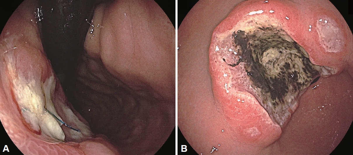
위 칸디다증(gastric candidiasis)

증 상

위 칸디다증의 임상 증상은 대부분 비특이적이며 무증상도 흔하다. 구역, 구토, 소화불량, 상복부 동통, 발열, 체중감소 및 상부 위장관 출혈에 이르기까지 다양하게 나타날 수 있다[8,31]. 면역 억제 상태나 종양 환자에서 호발한다.

진 단

양성 또는 악성 위궤양에 이차적으로 C. albicans가 침윤하여 발생하는 것이 보통이다[4,32,33]. 초기에는 홍반성 점막 변화가 관찰되며 점차 흰색 혹은 황색의 삼출성 위막이 형성되고, 궤양, 용종 및 점막 출혈이 관찰될 수 있다. 위 칸디다증을 내시경 소견에 따라 분류하면 발적된 점막 위에 백태가 덮힌 백태형(thrush type)과 일반적인 궤양과 차이가 없는 궤양형(ulcerated type), 결절형태를 취하는 결절형(nodular type)으로 분류할 수 있으며(Table 2 and Fig. 2), 이 중 백태형이 가장 빈도가 높다고 알려져 있다[34]. 대부분의 위 칸디다증에서는 다발성의 작은 궤양이나 미만성 표재성 미란이 관찰되지만 깊은 궤양으로도 진행할 수 있다. 칸디다 감염이 동반된 위궤양은 일반적인 위궤양보다 직경이 크고, 궤양저가 지저분하여 진행성 위암과 같은 악성 종양으로 오인될 수 있다[34]. 또한 궤양이 동반된 점막하병변 등으로 관찰되기도 하여 타겟 형태 또는 ‘bull’s eye’ 형태의 종양으로 보일 수도 있다[35]. 식도 칸디다증과 같이 조직병리학적 검사로 확진한다.

치 료

증상이 있는 위 칸디다증으로 의심되는 경우 플루코나졸 투여를 고려할 수 있으나 이에 대해서는 아직까지 정립된 의견은 없다. 칸디다의 궤양 침윤이 정상 궤양 치유 과정을 지연시키기 때문에 궤양 치유를 위하여 항진균제를 사용해야 한다는 의견과[36], 위궤양에서 같이 확인되는 진균 감염은 이차 감염이므로 궤양의 치료 및 자연 경과에 영향이 없으므로 항진균제를 사용할 필요가 없다는 의견도 있다[37]. 국내 후향적 연구에서 양성 위궤양에 동반된 칸디다 감염은 위산분비 억제제 투여만으로 궤양이 호전되며 함께 호전되는 경과를 취하므로 기회감염에 의한 것으로 보여 추가적인 항진균제 투약은 필요하지 않다고 보고하였다[38]. 칸디다 위궤양 환자에서 대부분 항진균제를 사용할 필요는 없겠으나, 고령, 암환자, 항암치료 중인 환자, 천공의 위험이 있는 경우, 전신 진균 감염 가능성이 있는 경우는 항진균제 중 폴리엔(polyene) 계열인 니스타틴, 암포테리신 B 등의 사용이 필요하다. 식도 칸디다증에 준하여 임상 증상의 호전을 보일 때까지 14-21일간 플루코나졸 200-400 mg 경구 투여가 선호된다. 경구 투여가 어려운 경우 플루코나졸 400 mg, 암포테리신 B 0.3-0.6 mg/kg, 에키노칸딘(echinocandin)계 항진균제를 정맥 투여하고, 플루코나졸에 효과가 없는 경우에는 이트라코나졸 200 mg, 포사코나졸(posaconazole) 800 mg, 보리코나졸(voriconazole) 400 mg 또는 암포테리신 B, 에키노칸딘계 항진균제로 대체하여 14-21일간 치료해 볼 수 있다[27]. 그러나 심한 면역억제 상태의 환자에서는 에키노칸딘계 항진균제 또는 암포테리신 B를 1차로 투여하는 것이 추천된다[27].

소장 칸디다증

십이지장을 포함한 소장에 발생하는 칸디다증은 매우 드물다. 국내 증례보고들은 만성 빈혈 환자에서 발생된 십이지장 칸디다증[39], C. albicans 감염에 의한 비전형적 거대 십이지장 궤양[40], 약물사용으로 인한 칸디다증을 동반한 무증상의 십이지장 궤양 환자에 대한 보고가 있다.

위장관 모균증(gastrointestinal mucormycosis)

모균증은 털곰팡이목(order Mucorales)에 의해 발생하는 치명적인 진균 감염으로, 털곰팡이증 혹은 검은 곰팡이증으로도 불리며, 과거에는 zygomycosis로도 불렸다. 모균증은 전체 진균 감염의 10%를 차지한다. 모균증은 코-안와-뇌 침범의 형태가 가장 흔하다. 위장관 침범은 드문 것으로 알려져 있으나 지난 20-30년 동안 위장관 모균증이 증가하는 추세로 보고되었다[41,42]. 침습성 모균증에서 위장관 침범은 7%-13%에서 확인되었으며, 이 중 위에서 58%, 나머지 42%는 소장 및 대장에서 진단되었다[43]. 위장관 모균증은 주로 면역 저하와 관련이 있으나41 정상 면역 환자에서도 보고된 바 있다[42,44]. 기존의 소화성 궤양 질환, Mucorales로 오염된 음식과 물의 섭취, 오염된 비위관, 설압자 및 나무 주걱의 사용은 모균증이 위장관에 침범할 가능성이 있는 요인으로 알려져 있다[45,46].

증 상

위장관 모균증 증례의 약 25%만이 임상적으로 진단되고, 위장관 모균증은 급성 경과와 급속한 치명적 특성으로 인해 부검에서 처음으로 발견되기도 한다[47]. 위장관 모균증은 발생 빈도가 낮고 복통, 팽만감, 발열, 설사 등의 비특이적인 증상으로 시작하여 위장관 출혈, 천공 및 급격한 활력징후의 악화 등 다양하게 나타나 임상적으로 조기에 진단하는 것은 매우 어렵다[42]. 모균은 혈관과 장벽을 침범하여 패혈증, 복막염, 대량 위장 출혈 및 장 천공을 일으키며, 장천공과 상부 위장관 출혈로 인한 사망률은 85%에 이른다[45].

진 단

위 모균증은 내시경에서 일반적으로 괴사가 동반된 경계가 뚜렷한 커다란 궤양의 형태로 관찰되며, 두껍게 부착된 녹색 삼출물이 동반된다[43]. 확진은 진균 배양검사를 통해 할 수 있다. 조직병리학적 검사에서 GMS 또는 PAS 염색을 시행하면 균사가 직각 또는 둔각으로 가지를 뻗는 격막 형태와 격벽이 없는 균사(6-25 μm)를 관찰할 수 있다[47]. 영상학적으로는 모균증에서 결절과 흉막 삼출이 흔하다고 알려져 있으나 이러한 소견들은 비특이적이다. 혈청 갈락토만난 검사도 모균증의 조기 진단에 도움이 된다.

치 료

암포테리신 B 지용성 제제 5-10 mg/kg 정맥 투여가 추천되며 사용 기간은 질병의 호전 경과에 따른다. 추가적으로 진균 감염의 부피를 줄이고 감염의 추가 확산을 막기위해 감염된 조직의 외과적 괴사조직 제거(debridement)를 조기에 적극적으로 고려해야 한다.

위장관 아스페르길루스증(gastrointestinal aspergillosis)

아스페르길루스는 환경에서 일반적으로 발견되는 분생포자(conidia)를 형성하는 진균의 일종으로, 대부분의 경우 흡입된 진균 분생포자는 상기도의 점액 섬모나 폐포 대식세포를 통해 제거된다. 아스페르길루스 감염을 일으키는 진균은 대부분 Aspergillus fumigatus, A. flavus, A. niger이다. 아스페르길루스증의 90%-98%는 폐를 침범한다. 위 아스페르길루스증은 매우 드물며, 독립적으로 발생하기도하나 일반적으로 파종성 아스페르길루스증의 일부로 발생한다[48].
위장관 아스페르길루스증은 면역 저하 환자에서 주로 발견되는 침습성 아스페르길루스증의 드문 임상 증상이다. 위장관은 파종 후 두 번째로 흔히 영향을 받는 부위로, 일반 인구에서 위장관 아스페르길루스증의 발생률이 낮기 때문에 일반적인 감별진단으로는 잘 고려되지 않는다[49]. 위험 요인에는 세포 독성 화학 요법, 장기간의 호중구감소증, 스테로이드 장기 복용, 장기 이식 및 후천성면역결핍증이 있으며, 면역저하가 없는 집중 치료실 환자에서의 호발 요인은 아직 알려지지 않았다. 아스페르길루스증의 사망률은 30%-70%로 보고된다[50].

증 상

위장관 증상으로는 복통(38%), 팽만, 설사(21%) 및 위장관 출혈(20%)이 흔하다. 이외 전신 증상에는 발열, 피로, 구토 뿐만 아니라 중추신경계, 피부과 및 폐 후유증(예: 정신 상태 변화, 마른 기침 및 부비동염)이 포함된다[49]. 아스페르길루스의 혈관 침습적 특성으로 혈관 혈전증을 유발하여 혈액성 설사 형태의 위장관 출혈로 나타나는 장 허혈이 발생할 수 있고, 장 폐색에 의한 급성 충수돌기염이나 장 천공에 의한 급성 복막염으로도 나타날 수 있다. 원발성 위장 침범은 거의 관찰되지 않았으며 사례 보고도 거의 없다.

진 단

아스페르길루스증의 병리학적 특징을 나타내는 영상학적 소견은 없다. 병리조직학적으로 예각 분기를 갖는 격벽 균사를 확인하는 것이 진단에 중요하다. 혈청 검사로 베타글루칸 또는 갈락토마난을 확인해볼 수 있으나 위장관 감염의 진단에 비특이적이다. 갈락토마난은 Penicillium , BotrytisPaecilomyces를 포함한 다른 곰팡이와의 교차 활성이 있고, Penicillium (피페라실린/타조박탐, 아목시실린-클라불라네이트)이 생산하는 항생제 사용으로도 위양성 결과가 나올 수 있다.

치 료

보리코나졸이 첫 번째로 선택된다.

결 론

이번 글에서는 진균에 의해 감염될 수 있는 상부위장관 질환에 대해 알아보았다. 상부위장관 진균 감염은 주로 면역 저하 상태에서 기회감염의 형태로 나타나고, 주로 칸디다에 의해 발생하며 이외 모균이나 아스페르길루스에 의해서도 감염될 수 있다. 칸디다에 의한 질환은 식도의 경우 치료 대상자를 선정하는 것이 중요하며, 위 칸디다의 경우 대개 항진균제 치료는 필요치 않고 궤양 치료만으로도 호전이 가능하다. 모균증이나 아스페르길루스증은 드문 질환이나 비특이적 임상양상으로 발현하여 급속도의 다양한 경과를 취할 수 있으므로 기저질환을 고려하여 질병을 조기에 의심하는 것이 중요하다.
결론적으로 면역저하자에서 비특이적 위장관 증상이 지속될 때는 진균 감염에 의한 상부위장관 질환 가능성에 대한 고려가 필요하다. 진균성 상부위장관 질환을 잘 이해하는 것은 질병을 조기에 진단하고 치료를 빨리 시작하여 환자의 예후를 향상시키는 데 도움이 될 것이다.

Notes

Availability of Data and Material

All data generated or analyzed during the study are included in this published article.

Conflicts of Interest

The authors have no financial conflicts of interest.

Funding Statement

None

Authors’ Contribution

Conceptualization: Ayoung Lee, Sung Woo Jung. Data curation: Ayoung Lee. Formal analysis: Ayoung Lee, Sung Woo Jung. Investigation: Ayoung Lee. Methodology: Ayoung Lee, Sung Woo Jung. Project administration: Ayoung Lee, Sung Woo Jung. Resources: Ayoung Lee, Sung Woo Jung. Software: Ayoung Lee. Supervision: Sung Woo Jung, Jung Mogg Kim. Validation: all authors. Visualization: Ayoung Lee, Sung Woo Jung. Writing—original draft: Ayoung Lee. Writing—review & editing: all authors. Approval of final manuscript: all authors.

Acknowledgements

None

Fig. 1.
Esophageal candidiasis. Endoscopic severity of Kodsi’s classification. A: Grade I, a few raised white plaques up to 2 mm in size without edema or ulceration. B: Grade II, multiple raised white plaques greater than 2 mm in size without ulceration. C: Grade III, confluent, linear, and nodular elevated plaques. D: Grade IV, finding of grade III with increased friability of the mucous membranes and occasional narrowing of the lumen.
kjhugr-2024-0029f1.jpg
Fig. 2.
Gastric candidiasis. Endoscopic severity of gastric candidiasis. A: Ulcerative type, exudate with ulcer. This patient underwent esophagectomy for esophageal cancer, and a benign gastric ulcer with candidiasis was identified below the anastomosis site. B: Gastric candidiasis with the ulcer base in a patient with gastric MALToma.
kjhugr-2024-0029f2.jpg
Table 1.
Kodsi’s classification
Grade Number of plaques Shape Size Associated finding
I A few Raised white plaques Up to 2 mm Without edema or ulceration
II Multiple Raised white plaques Greater than 2 mm Without ulceration
III Multiple Confluent, linear, and nodular elevated plaques Greater than 2 mm With ulceration
IV Multiple Confluent, linear, and nodular elevated plaques Greater than 2 mm With increased friability of the mucous membranes, occasional narrowing of the lumen
Table 2.
Gastric candidiasis
Thrush type Ulcerative type Nodular type
Feature White or green-white membrane, readily removed and spread over inflamed mucosa No particular endoscopic features Nodular projections with an apparent diameter of a few millimeters, overlaid by highly inflamed mucosa
Location Variable Variable Mainly antrum
Extension Variable Variable Variable

REFERENCES

1. Ost KS, Round JL. Commensal fungi in intestinal health and disease. Nat Rev Gastroenterol Hepatol 2023;20:723–734.
crossref pmid pdf
2. Lionakis MS, Edwards Jr JE. editors. Pathogenesis, diagnosis, and treatment of fungal infections. In: Loscalzo J, Fauci A, Kasper D, Hauser S, Longo D, Jameson JL, editors. Harrison’s principles of internal medicine, 21e. New York: McGraw-Hill Education, 2022.

3. Bongomin F, Gago S, Oladele RO, Denning DW. Global and multinational prevalence of fungal diseases-estimate precision. J Fungi (Basel) 2017;3:57.
crossref pmid pmc
4. Seo TJ, Lee WS, Lee KR, et al. Gastric candidiasis in an immunocompromised host: a case report and review of the literature. Korean J Gastrointest Endosc 2010;40:170–174.

5. Hallen-Adams HE, Suhr MJ. Fungi in the healthy human gastrointestinal tract. Virulence 2017;8:352–358.
crossref pmid
6. Kavyashree M, Pal B, Dutta S, Ashok Badhe B, Nelamangala Ramakrishnaiah VP. Gastric candidiasis leading to perforation: an unusual presentation. Cureus 2021;13:e17878.
crossref pmid pmc
7. Choi JH, Lee CG, Lim YJ, Kang HW, Lim CY, Choi JS. Prevalence and risk factors of esophageal candidiasis in healthy individuals: a single center experience in Korea. Yonsei Med J 2013;54:160–165.
crossref pmid
8. Kim ST, Kim HU, Lee DH. [Thick yellowish exudative pseudomembrane of the post-subtotal gastrectomy residual stomach]. Korean J Helicobacter Up Gastrointest Res 2022;22:240–242; Korean.
crossref pdf
9. Perlroth J, Choi B, Spellberg B. Nosocomial fungal infections: epidemiology, diagnosis, and treatment. Med Mycol 2007;45:321–346.
crossref pmid
10. Kothavade RJ, Kura MM, Valand AG, Panthaki MH. Candida tropicalis: its prevalence, pathogenicity and increasing resistance to fluconazole. J Med Microbiol 2010;59(Pt 8): 873–880.
crossref pmid
11. Jo H, Shin CM. Infectious gastric diseases other than Helicobacter. Korean J Gastroenterol 2023;82:269–281.
crossref pmid
12. Rajablou M, Ganz RA, Batts KP. Candida infection presenting as multiple ulcerated masses. Gastrointest Endosc 2007;65:164–166.
crossref pmid
13. Lee BE. [Differential diagnosis of esophageal ulcers]. Korean J Helicobacter Up Gastrointest Res 2023;23:68–72; Korean.
crossref pdf
14. Gock M, Schäfer M, Perren A, Demartines N, Clavien PA. Fatal esophageal perforation caused by invasive candidiasis. Ann Thorac Surg 2005;80:1120–1122.
crossref pmid
15. Dean DA, Burchard KW. Surgical perspective on invasive Candida infections. World J Surg 1998;22:127–134.
crossref pmid pdf
16. Kodsi BE, Wickremesinghe C, Kozinn PJ, Iswara K, Goldberg PK. Candida esophagitis: a prospective study of 27 cases. Gastroenterology 1976;71:715–719.
crossref pmid
17. Nishimura S, Nagata N, Shimbo T, et al. Factors associated with esophageal candidiasis and its endoscopic severity in the era of antiretroviral therapy. PLoS One 2013;8:e58217.
crossref pmid pmc
18. Young JA, Elias E. Gastro-oesophageal candidiasis: diagnosis by brush cytology. J Clin Pathol 1985;38:293–296.
crossref pmid pmc
19. Bougnoux ME, Hill C, Moissenet D, et al. Comparison of antibody, antigen, and metabolite assays for hospitalized patients with disseminated or peripheral candidiasis. J Clin Microbiol 1990;28:905–909.
crossref pmid pmc pdf
20. Lee SP, Sung IK, Kim JH, Lee SY, Park HS, Shim CS. The clinical course of asymptomatic esophageal candidiasis incidentally diagnosed in general health inspection. Scand J Gastroenterol 2015;50:1444–1450.
crossref pmid
21. Min YW, Kim E, Son HJ, Kim JJ, Rhee PL. Antifungal treatment is not required for immunocompetent individuals with asymptomatic esophageal candidiasis. Medicine (Baltimore) 2015;94:e1969.
crossref pmid pmc
22. Lionakis MS, Singh S, Ibrahim AS, Edwards Jr JE. Candidiasis. In: Loscalzo J, Fauci A, Kasper D, Hauser S, Longo D, Jameson JL, editors. Harrison’s principles of internal medicine, 21e. New York: McGraw-Hill Education, 2022.

23. LiverTox: clinical and research information on drug-induced liver injury. Bethesda: National Institute of Diabetes and Digestive and Kidney Diseases, 2012.

24. Wang JL, Chang CH, Young-Xu Y, Chan KA. Systematic review and meta-analysis of the tolerability and hepatotoxicity of antifungals in empirical and definitive therapy for invasive fungal infection. Antimicrob Agents Chemother 2010;54:2409–2419.
crossref pmid pmc pdf
25. Benitez LL, Carver PL. Adverse effects associated with long-term administration of azole antifungal agents. Drugs 2019;79:833–853.
crossref pmid pdf
26. Pfizer. Fluconazole (Diflucan) product package insert, 2024 [accessed on May 23, 2024]. Available from: https://labeling.pfizer.com/Show-Labeling.aspx?id=575.

27. Pappas PG, Kauffman CA, Andes DR, et al. Clinical practice guideline for the management of candidiasis: 2016 update by the Infectious Diseases Society of America. Clin Infect Dis 2016;62:e1–e50.
crossref pmid pdf
28. Lange D, Pavao JH, Wu J, Klausner M. Effect of a cola beverage on the bioavailability of itraconazole in the presence of H2 blockers. J Clin Pharmacol 1997;37:535–540.
crossref pmid
29. Jaruratanasirikul S, Sriwiriyajan S. Effect of omeprazole on the pharmacokinetics of itraconazole. Eur J Clin Pharmacol 1998;54:159–161.
crossref pmid pdf
30. Janssen Pharmaceuticals, Inc. Itraconazole (Sporanox) product package insert, 2023 [accessed on May 23, 2024]. Available from: https://www.janssenlabels.com/package-insert/product-monograph/prescribing-information/SPORANOX-Capsules-pi.pdf.

31. Park KB, Chang YW, Kim HJ, et al. A case of gastric candidiasis presented with massive gastric bleeding. Korean J Gastrointest Endosc 2000;20:41–45.

32. Seo GS, Kim JA, Kim YS, et al. A clinical analysis of gastric candidiasis. Korean J Gastrointest Endosc 1999;19:26–32.

33. Hirasaki S, Koide N, Ogawa H, Tsuji T. Benign gastric ulcer associated with Canidida infection in a healthy adult. J Gastroenterol 1999;34:688–693.
pmid
34. Minoli G, Terruzzi V, Butti G, Frigerio G, Rossini A. Gastric candidiasis: an endoscopic and histological study in 26 patients. Gastrointest Endosc 1982;28:59–61.
crossref pmid
35. Cronan J, Burrell M, Trepeta R. Aphthoid ulcerations in gastric candidiasis. Radiology 1980;134:607–611.
crossref pmid
36. Neeman A, Avidor I, Kadish U. Candidal infection of benign gastric ulcers in aged patients. Am J Gastroenterol 1981;75:211–213.
pmid
37. Wu CS, Wu SS, Chen PC. A prospective study of fungal infection of gastric ulcers: clinical significance and correlation with medical treatment. Gastrointest Endosc 1995;42:56–58.
crossref pmid
38. Jung MK, Jeon SW, Cho CM, et al. Treatment of gastric candidiasis in patients with gastric ulcer disease: are antifungal agents necessary? Gut Liver 2009;3:31–34.
crossref pmid pmc
39. Seo SO, Oh HJ, Kim KH, et al. A cases of duodenal candidiasis in the patient of chronic anemia. Korean J Med 2009;77(Suppl 2): 287–290.

40. Cho JY, Jung HK, Kim DY, Moon IH, Cho MS. A case of large duodenal ulcer associated with Candida albicans. Korean J Gastrointest Endosc 2003;26:154–157.

41. Spellberg B. Gastrointestinal mucormycosis: an evolving disease. Gastroenterol Hepatol (N Y) 2012;8:140–142.
pmid pmc
42. Huang H, Xie L, Zheng Z, et al. Mucormycosis-induced upper gastrointestinal ulcer perforation in immunocompetent patients: a report of two cases. BMC Gastroenterol 2021;21:311.
crossref pmid pmc pdf
43. Monte Junior ESD, Santos MELD, Ribeiro IB, et al. Rare and fatal gastrointestinal mucormycosis (zygomycosis) in a COVID-19 patient: a case report. Clin Endosc 2020;53:746–749.
crossref pmid pmc pdf
44. Albtoosh AS, Shaf’ei M, Al Hayek S, et al. A successfully treated gastric mucormycosis in an immunocompetent patient: case report and literature review. Clin Case Rep 2023;11:e7540.
crossref pmid pmc
45. Hameed T, Jain SK, Ansari FM, Nizam A, Dua A. Spontaneous gastric necrosis: a rare presentation of invasive mucormycosis in an immunocompetent adult. Case Rep Infect Dis 2020;2020:7514051.
crossref pmid pmc pdf
46. Maraví-Poma E, Rodríguez-Tudela JL, de Jalón JG, et al. Outbreak of gastric mucormycosis associated with the use of wooden tongue depressors in critically ill patients. Intensive Care Med 2004;30:724–728.
crossref pmid pdf
47. Bhowmik S, Jadav D, Aggarwal D, Shekhawat RS. Gastric mucormycosis. Autops Case Rep 2023;13:e2023421.
crossref pmid pmc
48. Kulkarni AA, Aruni A, Rastogi P, Rana S, Gupta R. Invasive aspergillosis causing gastric necrosis and perforation: a case report. JGH Open 2020;4:90–93.
crossref pmid pdf
49. Yelika SB, Tumati A, Denoya P. Intestinal aspergillosis: systematic review on patterns of clinical presentation and management. Surg Infect (Larchmt) 2021;22:326–333.
crossref pmid
50. Sulik-Tyszka B, Figiel W, Krawczyk M, Wróblewska M. Invasive aspergillosis of the stomach and co-infection with Candida krusei in a patient with terminal liver failure: a case report. Transplant Proc 2016;48:3149–3152.
crossref


Editorial Office
Lotte Gold Rose II Room 917, 31 Seolleung-ro 86-gil, Gangnam-gu, Seoul 06193, Korea
Tel: +82-2-717-5543    Fax: +82-2-565-9947    E-mail: helicojournal@smileml.com                

Copyright © 2024 by Korean College of Helicobacter and Upper Gastrointestinal Research.

Developed in M2PI

Close layer
prev next