Korean J Helicobacter  Up Gastrointest Res Search

CLOSE


Korean J Helicobacter  Up Gastrointest Res > Volume 24(2); 2024 > Article
위의 드문 세균성 감염

Abstract

An acidic environment, gastric emptying, and abundant blood supply inhibit bacterial infection of the stomach. Helicobacter pylori can evade these defense mechanisms and is a well-known etiological contributor to chronic gastritis. Non-H. pylori bacterial infections such as acute phlegmonous gastritis, gastric syphilis, gastric tuberculosis, and gastric actinomycosis are uncommon and owing to their nonspecific findings, are diagnostically challenging in patients without a high index of clinical suspicion. Predisposition to bacterial infection is attributable to an increase in the prevalence of medical conditions and factors that precipitate immunosuppression, in addition to high rates of gastric mucosal injury associated with endoscopic procedures. Gastric bacterial infection negatively affects patients’ quality of life, increases the socioeconomic burden, and may occasionally be fatal. Therefore, physicians should be familiar with the endoscopic features and clinical manifestations of non-H. pylori bacterial infections of the stomach.

서 론

위는 위 내 산성 환경, 풍부한 혈류와 위배출, 항균성 단백질 등의 방어기전으로 인해 병태생리적으로 세균 감염이 일어나기 어려운 장기이다[1,2]. 헬리코박터 파일로리(Helicobacter pylori , H. pylori)는 요소분해효소를 분비하여 위산으로부터 자신을 보호해 위 내 방어기전을 회피할 수 있어 세균성 위 감염의 주요 원인으로 잘 알려져 있다[3]. 위산으로부터 방어기전을 갖추지 않은 H. pylori 이외 대부분의 세균에 의한 위 감염은 매우 드물고, 내시경 소견도 특이적이지 않아 진단이 쉽지 않다[3,4]. 결과적으로 치료가 지연되거나 다른 악성질환과 감별을 위해 수술을 진행하는 경우도 있다[5]. 본 글에서는 H. pylori 이외 드물지만 국내에서 발생할 수 있는 4가지 세균성 위감염 질환인 급성 봉소염성 위염, 위매독, 위결핵, 위 방선균증의 내시경 소견 및 진단과 치료에 대해 기술하고자 한다.

본 론

급성 봉소염성 위염(acute phlegmonous gastritis)

급성 봉소염성 위염은 위 점막하층의 세균성 감염을 시작으로 위전층(transmural)에 위부종이나 괴사가 발생할 수 있는 드물지만 치명적인 질환이다[6,7]. 급성 봉소염성 위염은 1820년에 Cruveilhier 등에 의해 최초로 기술되었으며, 전 세계적으로는 500건 이상이 보고 되었다[8,9]. 급성 봉소염성 위염은 1919년 항생제가 발견되기 전에 보고된 시기에는 사망률이 약 92%에 달했으며, 항생제가 발견된 이후인 1973-2003년까지의 보고에서는 이전에 비해 많이 나아졌다고는 하지만 여전히 42%로 높은 사망률을 보인다[8]. 만성적인 음주력, 만성신부전, 조절 불량 상태의 당뇨, 면역저하 등의 위험 요인을 가진 환자에서 점막 손상 부위를 통한 세균 감염이 병의 기전으로 제시되고 있다[6-8,10,11]. 최근에 증가하고 있는 내시경 절제술로 인한 점막 결손뿐만 아니라 조직검사 또는 내시경 초음파(endoscopic ultrasonography)를 통한 미세침흡인검사(fine needle aspiration)와 같은 미세한 점막 손상 이후에도 급성 봉소염성 위염이 발생한 증례들이 보고된 바 있다[11-16]. 급성 봉소염성 위염의 주요 원인균으로는 Streptococci가 있으며, 전체 감염 중 약 70%-75%를 차지한다. 또한, Staphylococci , Escherichia coli , Haemophilus influenza , Proteus, Clostridia와 같은 균들도 위 생검 조직이나 위액 배양에서 확인될 수 있다[7,8,17]. 가스를 형성하는 균에 의한 감염의 경우 위벽에 공기가 형성되는 기종성 위염(emphysematous gastritis) 으로 나타날 수 있다[8]. 또한, 괴사성 위염(necrotizing gastritis)은 급성 봉소염성 위염의 가장 치명적인 형태로, 점막하 혈관에 광범위하게 형성되는 혈전이 특징이다[8]. 괴사성 위염은 위벽이 괴사되면서 복막염으로 빠르게 진행할 수 있어서, 진단이 지연되거나 간과될 경우 사망률이 높다[15,18].

임상 소견

환자는 일반적으로 복통, 구역, 구토 및 발열, 오한과 같은 감염의 징후가 관찰된다[6,7]. 질환이 진행되어 경과가 악화되면 복부 압통 및 반발통과 같은 복막 자극 징후가 분명해지고 화농성 구토를 보이기도 한다[6-8,19].

내시경 소견

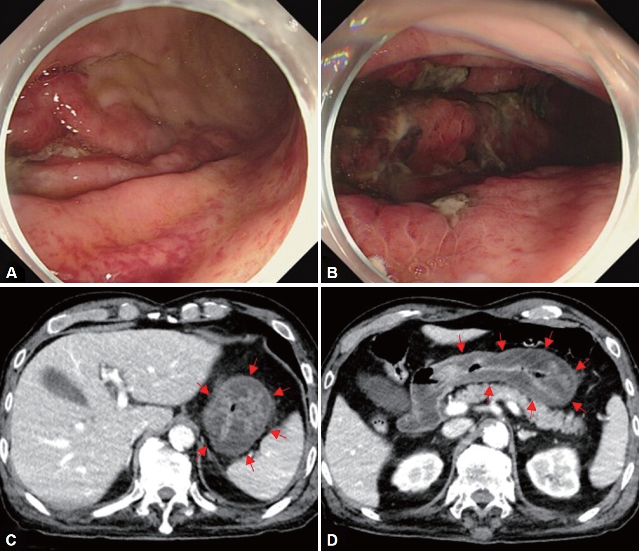
급성 봉소염성 위염은 침범 범위에 따라 국소형과 미만형으로 나눌 수 있는데, 국내 보고에서는 미만형이 더 흔하게 관찰되었다. 내시경 소견으로 점막 발적, 부종, 화농성 삼출물이 동반된 궤양이나 농양 등이 관찰될 수 있다(Fig. 1A and B). 위 전벽의 기능 저하로 위마비가 생기면 충분한 금식 기간에도 불구하고 저류된 음식물이 관찰될 수 있다[6].

영상 소견

복부 전산화 단층촬영(computed tomography)을 통하여 두껍고, 저음영의 위벽을 관찰할 수 있으며(Fig. 1C and D), 위벽 내 농양이 보이는 경우도 있다[6]. 초음파 내시경은 복부 전산화 단층촬영에 비해 위벽의 두께 및 염증파급정도를 평가하는데 유리하다[20]. 초음파 내시경에서 위벽이 미만성(diffuse) 으로 두꺼워져 있고, 저음영의 점막하층이 관찰될 수 있는데 이는 말트림프종(mucosa-assisted lymphoid tissue lymphoma) 초음파 내시경 소견과 비슷해 보일 수 있지만, 임상양상과 내시경 소견으로 감별이 가능하다[8].

진 단

복통 및 감염의 징후를 보이는 환자에서 내시경 소견 및 복부 전산화 단층촬영을 통해 의심해 볼 수 있고, 점막 및 점막하 생검 조직에 대해 그람 염색(gram stain) 및 세균 배양이 감염의 확진 및 원인균 동정에 도움이 된다[6,7,21]. 검사 시 주의사항으로 위점막 방어벽이 회복되지 않은 상태에서 내시경 검사를 하는 동안 주입된 공기로 인해 증가한 위내 압력이 혈관을 통한 세균의 역류를 유발해 패혈증(sepsis)을 일으킬 수 있다는 증례 보고가 있다. 따라서 급성 봉소염성 위염 환자에서 내시경을 시행할 때는 최소한의 공기 주입만 하고 검사 시간을 줄이려는 노력이 필요하다[14].

치료 및 예후

급성 봉소염성 위염은 1/3에서 복합균 감염이 발생하므로 초기 치료에는 3세대 세팔로스포린과 메트로니다졸 또는 피페라실린-타조박탐 또는 카바페넴 등의 광범위 항생제의 경험적 투여가 필요하다[8,17]. 중증의 환자가 아닌 경우 내과적 치료만으로 완치된 사례가 보고되었으며, 항생제 투여와 함께 내시경적 미세침흡인술을 통해 위벽 내 농양을 배농하여 수술 없이 완치된 사례도 있다[22]. 하지만 37명을 대상으로 한 보고에 따르면 질병이 미만성으로 파급된 환자 중에서 보존적인 치료를 받은 경우의 사망률은 60%였고, 오히려 수술적 치료를 받은 경우의 사망률은 33%였다[8]. 따라서 항생제 치료를 지속하는 중에 환자의 상태가 악화된다면 적극적인 수술적 치료를 고려하는 것이 필요하다[8].

위매독(gastric syphilis)

매독은 성매개감염으로 spirochete class에 속하는 Treponema pallidum에 의해 발생한다[23]. 국내에서는 2019년 매독 전수감시에서 1753명의 환자 발생이 보고되었으며, 이 중 남성은 1276명(72.9%), 여성은 474명(27.1%)이었다. 연령별로 20-40대가 1281명(73.2%)으로 많았으며, 60세 이상은 192명(11.0%)이었다. 병기별로 1기 매독은 1176명, 2기 매독은 554명, 선천성 매독은 23명 보고되었다[24]. 위매독은 보통 2기 매독에서 발생하며, 전체 매독 환자의 1% 미만에서 발생한다[25,26]. 52명의 위매독 환자를 분석한 보고에 따르면 남자가 많았으며(63%), 평균연령은 39세였고, 과거에 매독을 진단 받았던 경우는 13%에 불과했다[26]. 위매독은 매독 병력에 대한 문진만으로 배제할 수 없고, 임상 소견이 비특이적이어서 의심하지 않으면 진단하기 어렵다. 특히 내시경소견, 병리소견도 위암이나 림프종과 비슷한 양상으로 나타날 수 있어서 진단에 어려움이 있다[26-28].

임상 소견

위매독은 철저한 병력 청취를 통해 매독 병력, 성매개감염(sexually transmitted infection) 위험요인 등의 확인이 필요하다. 위매독의 증상 중에서는 상복부 통증이 가장 흔하며, 그 다음으로 오심과 구토 증상이 빈번하게 나타난다. 토혈, 흑색변, 빈혈과 같은 상부 위장관 출혈소견이나 조기 포만감, 체중감소가 나타나는 사례도 있었다[26,29]. 신체 검진에서는 33%의 환자에서만 매독 징후가 관찰되었으며, 발진(65%), 림프절 병증(53%), 성기 궤양(35%) 순으로 빈도가 높았다[26]. 일부 환자에서만 매독 징후를 발견할 수 있기 때문에 정상적인 신체진찰만으로 위매독을 배제할 수 없다[5,30].

내시경 소견

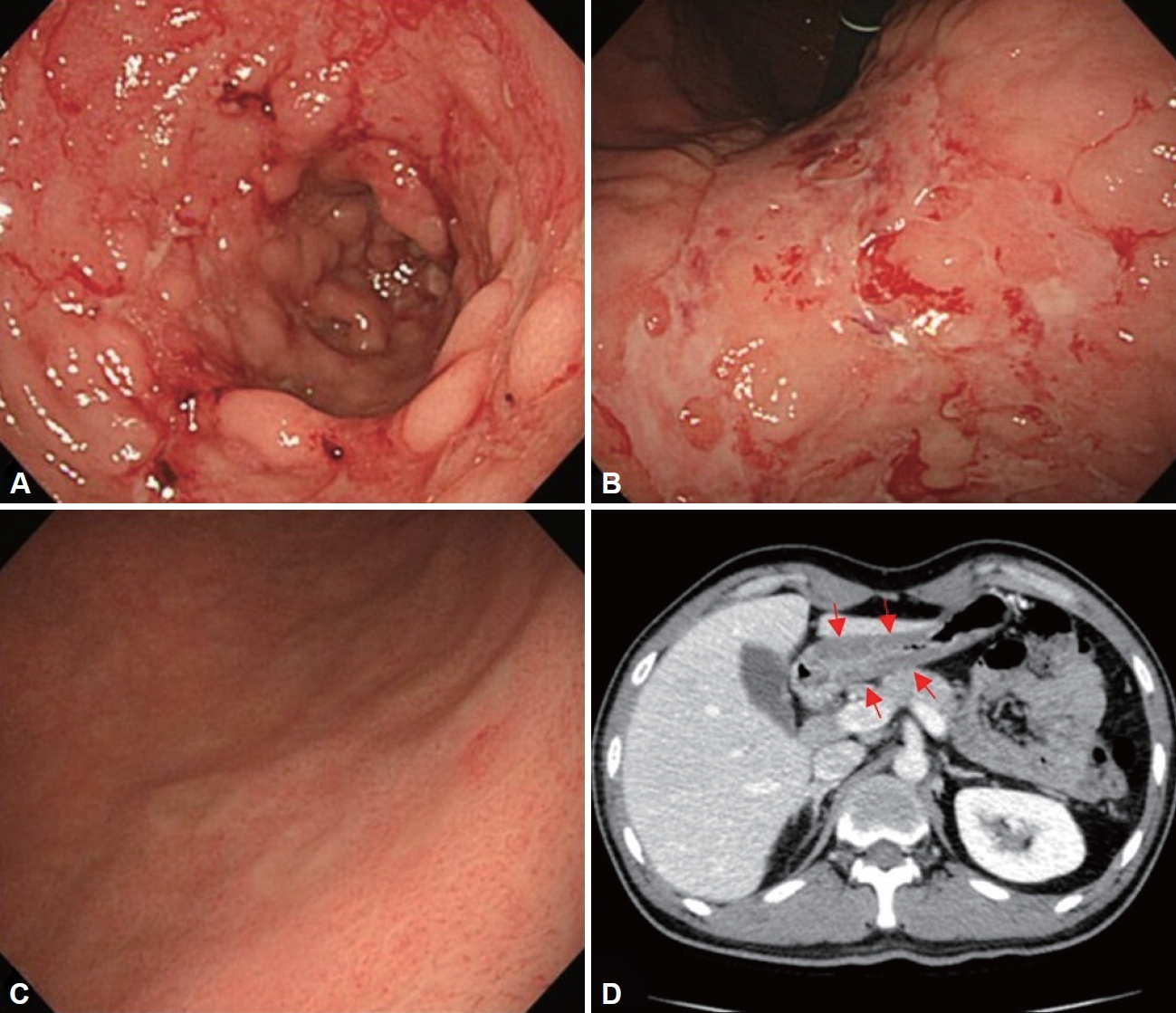
위매독의 주된 호발 부위는 전정부와 위각부, 원위부 체부이며, 상대적으로 위 기저부나 십이지장은 잘 침범하지 않는다[26,31]. 내시경 소견으로는 다발성 궤양, 거대 궤양, 미란, 두꺼워진 점막 주름, 결절성(nodular) 점막, 종괴(mass), 협착(stenosis)의 형태를 보일 수 있으며, 여러 형태의 병변이 혼재되어 있는 경우가 많다(Fig. 2A and B) [5]. 동전 모양의 구진(papule) 이 나타나면 위매독을 가장 우선적으로 고려해 볼 수 있다(Fig. 2C). 내시경적으로 결핵, 크론병(crohn’s disease), 사르코이드증(sarcoidosis), 보만 4형 위암(borrmann type 4), 림프종, 호산구성 위염(eosinophilic gastritis) 등과 비슷한 형태를 보일 수 있다[26,29,32,33]. 제산제 치료에도 치유되지 않는 궤양(refractory ulcer)이나 비전형적인 점막 변형을 가진 병변에서 진단이 안될 때 위매독을 의심을 해볼 수 있다[5].

영상 소견

복부 전산화 단층촬영에서 위벽의 협착, 점막주름의 불규칙적인 비대가 관찰되며(Fig. 2D), 체부에 경직(stiffness)이 진행되면서 모래시계(hourglass) 형태의 위가 나타난 보고도 있다[26,31].

진 단

매독의 혈청학적 검사로는 두 종류, 비트레포네마 검사(non-treponemal test)와 트레포네마 검사(treponemal test) 가 있다. 한 가지 종류의 검사만 시행한다면 위양성(falsepositive) 결과를 포함하여 최근 감염과 오래된 감염을 구별할 수 없기 때문에 두 가지 종류의 검사를 모두 시행한다. 선별검사로 주로 사용되는 비트레포네마 혈청학적 검사에는 rapid plasma regain test, venereal disease research laboratory 검사가 있고 지질 항원에 대한 항체를 측정하는 방법이다. 양성 검사 결과는 정량적 역가로 보고되며, 이는 질병의 활성도와 관련이 있다. 트레포네마 혈청학적 검사에는 fluorescent treponemal antibody absorption, T. pallidium latex agglutination, enzyme immunoassay 검사법 등이 있다. IgM은 후기 매독이나 치료받은 매독에서는 주로 음성이며, IgG는 치료받은 매독에서도 평생 양성이다. 매독에 대한 트레포네마와 비트레포네마 검사 모두에서 위양성 반응이 나타날 수도 있다. 위양성 결과의 가장 일반적인 원인은 고령, 자가면역질환, 심혈관계 질환, 임신, 말라리아, 다른 spirochete 감염 및 최근의 예방접종이 있다. 확진을 위해서는 위병변의 삼출물 또는 조직검사에서 트레포네마균을 검출하는 것이 필요한데, 은염색을 통해 spirochete를 확인할 수 있다[34]. 직접 형광항체검사(direct immunofluorescence staining)를 통해 검체에서 트레포네마 항원을 검출할 수 있는 방법은 민감도, 특이도가 높고 비교적 값이 싸면서 결과가 48시간 내에 나오는 장점이 있다[5]. 진단적 민감도를 향상시킬 목적으로 실시간 중합효소연쇄반응검사(real-time polymerase chain reaction) 를 사용할 수 있지만, 20% 정도의 위양성률이 함께 보고되고 있어 해석에 주의가 필요하다. 위매독 병변 조직의 haematoxylin-eosin (H&E) 염색 시 대부분 플라즈마 세포나(plasma cell), 림프구의 침윤으로 인한 미만성 위염이 관찰되며 궤양이 동반될 수 있다. 혈관염은 매독 침범의 특징적인 소견이지만, 혈관이 위치하는 점막하층 검체가 필요하다. 이를 위해 내시경적 점막 절제술(endoscopic mucosal resection) 이나 탐색적 개복술(explorative laparotomy)을 통해 위 매독을 진단한 사례 보고도 있다[5]. 조직검체에서 림프상피병변(lymphoepithelial lesion)과 상피세포 핵의 비전형(nuclear atypia)을 보이는 경우에는 위암이나 위림프종으로 오인할 수 있다[28]. 조직검사에서도 확진이 어려운 경우 혈청학적 검사 소견을 확인하고 경험적으로 항생제를 투여한 후, 단기 추적 관찰하면서 증상 호전 및 내시경적 병변 소실 여부를 종합해서 진단할 수 있다[4].

치료 및 경과

성인 1기, 2기 매독의 치료는 벤자신 페니실린G (benzathine penicillin G) 240만 단위를 1회 근육 내 투여하는 것이다[30]. 보고에 따르면 치료 성공률은 90% 정도이며, 임상 증상이 가장 빠르게 좋아져 보통 3-4일 후 호전된다. 내시경 소견에서 궤양이나 미란은 치료 시작 후 10일 정도면 호전되지 만 점막 비후와 위벽 경직과 같은 소견은 오랜 시일 이후에 호전되는 것으로 보고된다[23]. 천공이나 폐쇄와 같은 합병증은 드물게 보고되었으며, 악성종양이나 림프종으로 오인하여 수술한 사례도 있다[5].

위결핵(gastric tuberculosis)

폐결핵 환자 0.5% 미만에서 속발성으로 위결핵이 발생할 수 있으며, 결핵균의 섭취로 인하여 직접적인 점막의 감염이 일어나거나, 혈행성 확산(hematogenous spread), 복강 내 임파절을 포함한 주변 장기로부터 전파로 인해 발생한다. 폐 또는 다른 위장관 침범 없이 위에서만 발생한 사례도 있으나 매우 드물다[35,36].
우리나라와 같이 결핵 유병률이 아직 높은 지역에서는 면역억제자가 아니라 하더라도 내시경 소견상 미만성의 침윤성(infiltrative) 병변이나 불응성 궤양, 위출구 협착, 폐쇄 등이 관찰되면 위결핵을 고려해야 한다[37].

임상 증상

위결핵 환자들은 대부분 결핵의 일반적인 특징인 발열 및 체중감소가 동반된다. 상복부 통증 및 위장관 출혈을 호소할 수도 있으며, 위 전정부와 유문 침범이 있거나 결핵성 림프절 등에 의한 외부 압박이 발생하는 경우 구역 및 구토와 같은 위출구 폐쇄증상을 보이기도 한다[38-40].

내시경 소견

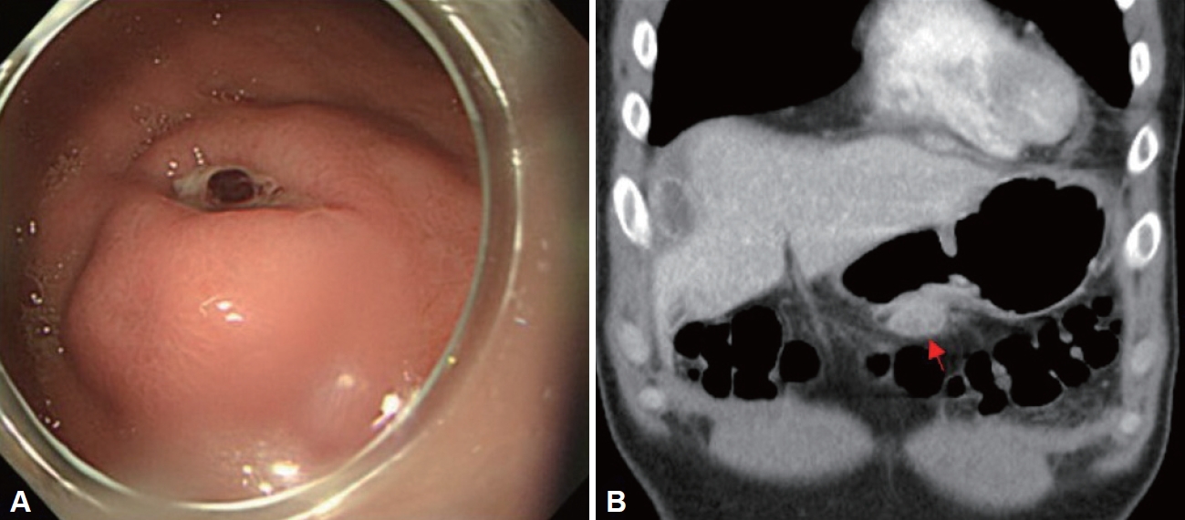
위결핵의 내시경 소견은 비특이적이며 점막의 비후나 발적, 불규칙적인 미란, 궤양 등으로 관찰될 수 있다. 따라서 이러한 형태의 병변을 보일 수 있는 소화성 궤양, 크론병, 매독, 림프종, 사르코이드증 등과 감별이 필요하다. 또한 위점막하 종양(submucosal tumor)과 유사한 형태로 나타날 경우 위점막하 유사 위암이나 위간질성종양(gastrointestinal stromal tumor)과 감별이 필요하다(Fig. 3A) [41]. 위 전층으로 염증이 심해지면 담관 또는 다른 위장관으로 누공(fistula)이 생기기도 한다[42].

영상 소견

위결핵은 복부 전산화 단층촬영에서 두꺼워진 위벽이나 위점막하 종양의 형태로 관찰되며(Fig. 3B), 종종 국소 림프절 비대가 보인다[43]. 폐결핵이 동반되는 경우가 많기 때문에 반드시 흉부 X선 촬영 결과를 확인하고 필요하다면 흉부 전산화 단층촬영도 적극적으로 사용해야 한다[44].

진 단

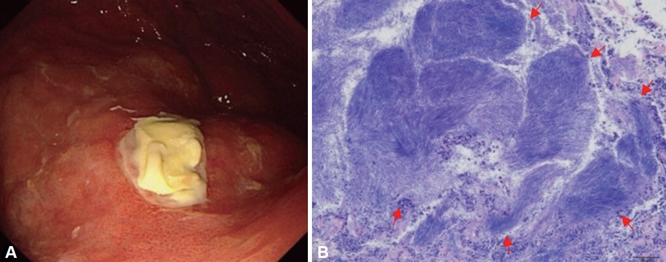
조직 검체의 Ziehl-Neelsen 염색에서 막대 모양의 항산균(acid-fast bacilli)이 염색되거나 건락화된 육아종(caseating granuloma) 소견 또는 랑게르한스 거대세포(Langerhans giant cell)가 확인되면 위결핵으로 진단할 수 있다. 하지만 전형적인 결핵 소견이 나타나지 않는 경우가 많으며, 특히 병변이 점막하층에 깊게 위치하는 경우 육아종 확인 및 항산균 발견이 어려운 경우가 있다. 따라서 충분한 조직검사와 함께 조직을 통한 균 배양과 결핵 중합효소연쇄반응검사를 함께 진행하는 것이 추천된다[43].

치 료

결핵의 치료는 6개월 표준 항결핵제 복용이 원칙이다. 초기 2 개월은 집중치료기로 이소니아지드, 리팜핀, 에탐부톨, 피라진아미드를 동시에 복용한다. 이어지는 4개월 유지치료기에는 이소니아지드, 리팜핀, 에탐부톨을 같이 복용한다[45]. 약물치료로 호전되지 않는 궤양이나, 폐쇄, 농양, 누공 등의 합병증이 발생한다면 위절제 및 우회술(bypass) 등 수술적 치료를 고려해야 한다[43]. 또한, 협착이 발생한 경우 풍선확장술이 효과적이었다는 보고가 있다[46].

위 방선균증(gastric actinomycosis)

위 방선균증은 구강, 위장관, 비뇨생식기관에 상재하는 그람양성이며, 혐기성 균인 Actinomyces israelii에 의해 발생하는 만성 화농성, 육아종성 질환이다[47]. Actinomyces israelii는 손상된 점막에 침투하여 화농성 염증 반응을 유발하며, 결과적으로 농양 및 농루를 형성한다. 위는 위산으로 인해 방선균의 침입과 성장을 막기 때문에 원발적인 위 방선균증은 매우 드물고 수술 후 혹은 고령의 장기간 제산제 복용자, 면역억제자 등에서 발생한다[48,49].

임상 증상

위 방선균증의 임상 소견은 비특이적이며, 무증상이거나 상복부 통증, 체중 감소, 위장관 출혈, 발열 그리고 드물게 위 출구 폐쇄가 나타날 수 있다[50-53].

내시경 소견

현재까지 상피하 종양 형태나 침윤 병변 혹은 궤양 형태로 관찰된 증례 보고들이 있다(Fig. 4A) [49,52-55]. 내시경 초음파에선 비대해진 위벽 내 불균질한 저에코성과 무에코성 병변으로 관찰되었다는 보고가 있다[49,56].

영상 소견

특징적인 영상학적 소견은 없지만 복부 전산화 단층촬영에서는 전반적으로 두꺼워진 위벽에 동반된 침윤된 병변으로 관찰되기 때문에 위암이나 림프종으로 오인되기도 한다[55]. 따라서 임상에서 진단은 수술 후 병리 결과에서 진단되는 경우가 많다[49,53,54,57].

진 단

위 방선균증의 확진은 균 배양 및 조직 검사를 통해 이루어지는데, 혐기성 배양이 어렵고 3주까지도 걸릴 수 있기 때문에 주로 특징적인 조직 소견을 통해 진단이 된다. 특징적인 소견으로 H&E 염색에서 방선균이 서로 붙어서 가는 실 모양으로 모여 황과립(sulfur granule)을 형성하는 것이 관찰된다(Fig. 4B) [48].
그러나 염증이 침윤성으로 점막하층 밑으로 진행하면, 내시경 겸자생검에서는 비특이적인 염증 소견만 나타날 수 있다[49].

치 료

일반적으로 방선균은 적절한 치료에 좋은 예후를 가지는데, 베타락탐계열 항생제에 감수성이 높기 때문에 1차 치료로 페니실린G나 아목시실린을 사용한다. 하지만 상당수에서 이차성 섬유화를 동반하므로 경구 항생제를 포함해서 6-12개월 장기 치료가 필요하다[48,49,58]. 감염된 조직 절제와 적절한 항생제 치료로 치료 기간을 3개월로 줄일 수 있다[58].

결 론

H. pylori 이외 위의 세균성 감염은 드물고, 비특이적인 임상소견을 보이기 때문에 의심하지 않으면 진단하기 어렵다. 따라서 비특이적이더라도 강도가 심한 위장관 증상을 호소하는 환자는 상부 내시경 검사 전 면역 상태에 영향을 줄 수 있는 환자의 임상 정보 등을 적극적으로 파악하고 검사를 시행해야 한다. 또한 진단을 내리기 어려운 비전형적인 내시경 및 임상양상을 가진 경우에는 다시 충분한 병력 청취와 신체 진찰을 해야 한다. 그리고 내시경 및 영상소견, 혈청학적 검사, 병리검사에 대해서 다시 검토하고 마지막으로 치료에 대한 반응을 종합해 최종적으로 진단해야겠다. 본고에서 서술한 H. pylori 이외 세균성 위감염의 임상적 특징을 표로 비교해보면 다음과 같다(Table 1). 진단 지연은 환자의 삶의 질 저하, 사회비용적인 부담을 높일 뿐 아니라 때로는 치명적인 결과를 낳을 수 있기 때문에 본고에서 언급한 질병들에 대해서 숙지해야겠다.

Notes

Availability of Data and Material

Data sharing not applicable to this article as no datasets were generated or analyzed during the study.

Conflicts of Interest

The authors have no financial conflicts of interest.

Funding Statement

This study was supported by a 2024 research grant from Pusan National University Yangsan Hospital.

Authors’ Contribution

Conceptualization: Su Jin Kim. Investigation: Hyun Myung Cho. Resources: Jin Ook Jang. Supervision: Su Jin Kim. Validation: Jung Wook Lee. Visualization: Jin Ook Jang. Writing—original draft: Hyun Myung Cho. Writing—review & editing: Su Jin Kim. Approval of final manuscript: all authors.

Acknowledgements

We are truly thankful for sharing such a great case of gastric actinomycosis by Jung Wook Kim from Kyung Hee University.

Fig. 1.
Endoscopic and radiologic features of acute phlegmonous gastritis. A: Diffuse mucosal edema, loss of vasculature, erythema are shown. B: Erosions and ulcers are covered with purulent exudate, particularly prevalent on the greater curvature of the stomach. C and D: Abdominal computed tomography showed diffuse wall thickening of the stomach fundus and body (red arrows).
kjhugr-2024-0015f1.jpg
Fig. 2.
Clinical features of gastric syphilis. A and B: Mucosal thickening with nodular appearance, multiple erosions and large ulcers are located from the lesser curvature of the upper body to the antrum sparing the duodenum. C: Additionally, multiple whitish flat round papules were visualized at the body. D: Abdominal computed tomography revealed ulceroinfiltrating lesion from the body to the antrum (red arrows).
kjhugr-2024-0015f2.jpg
Fig. 3.
Endoscopic and radiologic findings of gastric tuberculosis. A: At the greater curvature of the stomach body, 3 cm sized submucosal tumor like mass with visible vessel in the center is observed. B: Abdominal computed tomography shows subepithelial mass (red arrow) at the greater curvature of the stomach body.
kjhugr-2024-0015f3.jpg
Fig. 4.
Endoscopic and histologic features of gastric actinomycosis. A: Endoscopic examination revealed 1 cm sized round ulcers, located on the anterior wall of the lower body and the posterior wall of the upper body. The ulcers were covered with yellowish exudate. B: Biopsy results showed chronic suppurative inflammation with sulfur granules. Sulfur granules were composed of basophilic filamentous bacterial aggregates (red arrows) surrounded by neutrophils (hematoxylin-eosin stain, ×100).
kjhugr-2024-0015f4.jpg
Table 1.
Summary of gastric infection caused by bacteria other than Helicobacter pylori
Acute phlegmonous gastritis Gastric syphilis Gastric tuberculosis Gastric actinomycosis
Predisposing factor Immunocompromised condition, mucosal injury Risk factors for sex-transmitted disease Pulmonary tuberculosis Previous gastric operation, immunosuppressed state
Clinical manifestation Epigastric pain, fever Epigastric pain, nausea/vomiting, occasionally rash, lymphadenopathy, genital ulcer Systemic manifestations of tuberculosis, nausea, vomiting Asymptomatic, epigastric pain, weight loss
Endoscopic findings Mucosal erythema/edema, ulcer, purulent exudate on body, gastroparesis Flat round papules, nodular mucosa, multiple erosions/ulcers on distal body, antrum Diffuse infiltration, ulcer, SMT like lesion, gastric outlet stenosis/obstruction Diffuse infiltration, ulcer, SMT like lesion
Radiologic findings Diffuse, hypodense thickening of body Irregular thickening of folds, narrowing, hourglass appearance Gastric wall thickening, regional lymphadenopathy Diffuse infiltrative gastric wall thickening
Diagnosis Gram stain, culture IFA, PCR, antibiotic response with serologic evidence Acid-fast bacilli on Ziehl-Neelsen stain, caseating granuloma, Langerhans giant cell, culture, PCR Sulfur granule, anaerobic culture

SMT, submucosal tumor; IFA, immunofluorescence assay; PCR, polymerase chain reaction.

REFERENCES

1. Nakamura K, Sakuragi N, Takakuwa A, Ayabe T. Paneth cell α-defensins and enteric microbiota in health and disease. Biosci Microbiota Food Health 2016;35:57–67.
crossref pmid
2. Lee SM, Cheung DY. Infectious diseases of the stomach in immunecompromised patients. Korean J Helicobacter Up Gastrointest Res 2019;19:38–41.
crossref pdf
3. Jo H, Shin CM. Infectious gastric diseases other than Helicobacter. Korean J Gastroenterol 2023;82:269–281.
crossref pmid
4. Roh M, Min YW. Infectious diseases of the upper gastrointestinal tract. Korean J Helicobacter Up Gastrointest Res 2019;19:16–22.
crossref pdf
5. Sinagra E, Macaione I, Stella M, et al. Gastric syphilis presenting as a nodal inflammatory pseudotumor mimicking a neoplasm: don’t forget the treponema! Case report and scoping review of the literature of the last 65 years. Gastroenterol Insights 2023;14:178–190.
crossref
6. Kim NY, Park JS, Lee KJ, Yun HK, Kim JS. A case of acute phlegmonous gastritis causing gastroparesis and cured with medical treatment alone. Korean J Gastroenterol 2011;57:309–314.
crossref pmid
7. Miller AI, Smith B, Rogers AI. Phlegmonous gastritis. Gastroenterology 1975;68:231–238.
crossref pmid
8. Kim GY, Ward J, Henessey B, et al. Phlegmonous gastritis: case report and review. Gastrointest Endosc 2005;61:168–174.
crossref pmid
9. Wakayama T, Watanabe H, Ishizaki Y, et al. A case of phlegmonous esophagitis associated with diffuse phlegmonous gastritis. Am J Gastroenterol 1994;89:804–806.
pmid
10. Joko T, Tanaka H, Hirakata H, et al. Phlegmonous gastritis in a haemodialysis patient with secondary amyloidosis. Nephrol Dial Transplant 1999;14:196–198.
crossref pmid
11. Lee BS, Kim SM, Seong JK, et al. Phlegmonous gastritis after endoscopic mucosal resection. Endoscopy 2005;37:490–493.
crossref pmid
12. Bron BA, Deyhle P, Pelloni S, Krejs GJ, Siebenmann RE, Blum AL. Phlegmonous gastritis diagnosed by endoscopic snare biopsy. Am J Dig Dis 1977;22:729–733.
crossref pmid pdf
13. Itonaga M, Ueda K, Ichinose M. Phlegmonous gastritis caused by endoscopic ultrasound-guided fine-needle aspiration (EUS-FNA). Dig Endosc 2012;24:488.
pmid
14. Kim TU, Kim SJ, Ryu H, et al. Emphysematous gastritis worsened after upper endoscopy. Korean J Helicobacter Up Gastrointest Res 2020;20:73–76.
crossref pdf
15. Kim KH, Kim CG, Kim YW, et al. Phlegmonous gastritis with early gastric cancer. J Gastric Cancer 2016;16:195–199.
crossref pmid pmc pdf
16. Ajibe H, Osawa H, Yoshizawa M, et al. Phlegmonous gastritis after endoscopic submucosal dissection for early gastric cancer. Therap Adv Gastroenterol 2008;1:91–95.
crossref pmid pmc pdf
17. Lee TH, Lee GS, Im EH, et al. A case of acute phlegmonous gastritis treated with antibiotics alone. Clin Endosc 2005;31:44–48.

18. Min SY, Kim YH, Park WS. Acute phlegmonous gastritis complicated by delayed perforation. World J Gastroenterol 2014;20:3383–3387.
crossref pmid pmc
19. Lee GW, Lee OJ, Jung KW, et al. Acute phlegmonous gastritis diagnosed early endoscopically and treated successfully with antibiotics. Clin Endosc 2001;23:225–229.

20. Iwakiri Y, Kabemura T, Yasuda D, et al. A case of acute phlegmonous gastritis successfully treated with antibiotics. J Clin Gastroenterol 1999;28:175–177.
crossref pmid
21. Kim HS, Jang WI, Lee SS, et al. Two cases of phlegmonous gastritis secondary to corrosive gastitis caused by formalin. Korean J Med 1991;40:268–273.

22. Kan-no Y, Irisawa A, Takagi T, et al. Endosonographic diagnosis and follow-up of phlegmonous gastritis. J Clin Ultrasound 2007;35:524–526.
crossref pmid
23. Greenstein DB, Wilcox CM, Schwartz DA. Gastric syphilis. Report of seven cases and review of the literature. J Clin Gastroenterol 1994;18:4–9.
pmid
24. Kim T, Cho SS, Kim HS, Choi H. National notification trend of sexually transmitted infections (STIs) in 2019-2020. Public Health Wkly Rep 2021;14:2496–2504.

25. Cooley RN, Childers JH. Acquired syphilis of the stomach. Report of two cases. Gastroenterology 1960;39:201–207.
crossref pmid
26. Mylona EE, Baraboutis IG, Papastamopoulos V, et al. Gastric syphilis: a systematic review of published cases of the last 50 years. Sex Transm Dis 2010;37:177–183.
crossref pmid
27. Abdu RA, Carter K, Pomidor WJ. Gastric syphilis mimicking linitis plastica. Arch Surg 1993;128:103–104.
crossref pmid
28. Long BW, Johnston JH, Wetzel W, Flowers RH 3rd, Haick A. Gastric syphilis: endoscopic and histological features mimicking lymphoma. Am J Gastroenterol 1995;90:1504–1507.
pmid
29. Yu HJ, Kim SJ, Oh HH, et al. Case report of gastric syphilis in Korea: clinical features, pathology, management, and prognosis. Medicine (Baltimore) 2021;100:e28212.
pmid pmc
30. Workowski KA, Bachmann LH, Chan PA, et al. Sexually transmitted infections treatment guidelines, 2021. MMWR Recomm Rep 2021;70:1–187.
crossref
31. Jones BV, Lichtenstein JE. Gastric syphilis: radiologic findings. AJR Am J Roentgenol 1993;160:59–61.
crossref pmid
32. Shen Y, Nie L, Zhang M, et al. Gastric syphilis mimicking lymphoma. Endoscopy 2015;47(Suppl 1): E170–E171.
crossref pmid
33. Massironi S, Carmagnola S, Penagini R, Conte D. Gastric involvement in a patient with secondary syphilis. Dig Liver Dis 2005;37:368–371.
crossref pmid
34. Ratnam S. The laboratory diagnosis of syphilis. Can J Infect Dis Med Microbiol 2005;16:45–51.
crossref pmid pmc pdf
35. Misra RC, Agarwal SK, Prakash P, Saha MM, Gupta PS. Gastric tuberculosis. Endoscopy 1982;14:235–237.
crossref pmid
36. Perez-Piqueras J, Coca S, Silva C, Martinez D, Peralba J, Moreno M. Isolated gastric tuberculosis: a case report. Endoscopy 1993;25:376.
crossref pmid
37. Kim SE, Shim KN, Yoon SJ, et al. A case of gastric tuberculosis mimicking advanced gastric cancer. Korean J Intern Med 2006;21:62–67.
crossref pmid pmc
38. Ecka RS, Wani ZA, Sharma M. Gastric tuberculosis with outlet obstruction: a case report presenting with a mass lesion. Case Rep Med 2013;2013:169051.
crossref pmid pmc pdf
39. Flores HB, Zano F, Ang EL, Estanislao N. Duodenal tuberculosis presenting as gastric outlet obstruction: a case report. World J Gastrointest Endosc 2011;3:16–19.
crossref pmid pmc
40. Kalpande S, Pandya JS, Tiwari A, Adhikari D. Gastric outlet obstruction: an unusual case of primary duodenal tuberculosis. BMJ Case Rep 2017;2017:bcr2016217966.
crossref pmid pmc
41. Kim TU, Kim SJ, Ryu H, et al. Gastric tuberculosis presenting as a subepithelial mass: a rare cause of gastrointestinal bleeding. Korean J Gastroenterol 2018;72:304–307.
crossref pmid pdf
42. Gilinsky NH, Marks IN, Kottler RE, Price SK. Abdominal tuberculosis. A 10-year review. S Afr Med J 1983;64:849–857.
pmid
43. Rao YG, Pande GK, Sahni P, Chattopadhyay TK. Gastroduodenal tuberculosis management guidelines, based on a large experience and a review of the literature. Can J Surg 2004;47:364–368.
pmid pmc
44. Elterefi AE, Uwaydah AK, Helal GR, Hassan NMM. Gastric tuberculosis presenting as a large gastric ulcer. BMJ Case Rep 2022;15:e248215.
crossref pmid pmc
45. World Health Organization. Comprehensive TB guidelines for national tuberculosis program. 5th ed. Geneva: World Health Organization, 2020.

46. Anand BS, Nanda R, Sachdev GK. Response of tuberculous stricture to antituberculous treatment. Gut 1988;29:62–69.
crossref pmid pmc
47. Scheckler WE. Observations on Wisconsin’s public health agenda. Wis Med J 1991;90:27, 29, 30–31.
pmid
48. Wong VK, Turmezei TD, Weston VC. Actinomycosis. BMJ 2011;343:d6099.
crossref pmid
49. Zumu M, Lakshmanan I, Arun RS, Damle A. Primary gastric actinomycosis: case reports and literature review. Gastroenterol Hepatol Endosc Pract 2024;4:34–39.
crossref
50. Lee YM, Law WL, Chu KW. Abdominal actinomycosis. ANZ J Surg 2001;71:261–263.
crossref pmid pdf
51. Choi MM, Baek JH, Lee JN, Park S, Lee WS. Clinical features of abdominopelvic actinomycosis: report of twenty cases and literature review. Yonsei Med J 2009;50:555–559.
crossref pmid pmc
52. Park SH, Song CW, Kim YB, Kim YS, Chun HR, Jeon HM. A case of asymptomatic primary gastric actinomycosis diagnosed following gastroscopic biopsy. Dig Dis Sci 2024;69:1071–1072.
crossref pmid pdf
53. Lee DS, Kang JY, Kim H, et al. A case of primary gastric actinomycosis. Korean J Med 2009;77:27–30.

54. Skoutelis A, Panagopoulos C, Kalfarentzos F, Bassaris H. Intramural gastric actinomycosis. South Med J 1995;88:647–650.
crossref pmid
55. Al-Obaidy K, Alruwaii F, Al Nemer A, Alsulaiman R, Alruwaii Z, Shawarby MA. Primary gastric actinomycosis: report of a case diagnosed in a gastroscopic biopsy. BMC Clin Pathol 2015;15:2.
crossref pmid pmc pdf
56. Lee SH, Kim HJ, Kim HJ, et al. Primary gastric actinomycosis diagnosed by endoscopic biopsy: case report. Gastrointest Endosc 2004;59:586–589.
crossref pmid
57. Euanorasetr C, Sornmayura P. Gastric outlet obstruction secondary to gastric actinomycosis: a case report and literature review. Thai J Surg 2010;31:63–68.

58. Valour F, Sénéchal A, Dupieux C, et al. Actinomycosis: etiology, clinical features, diagnosis, treatment, and management. Infect Drug Resist 2014;7:183–197.
pmid pmc


Editorial Office
Lotte Gold Rose II Room 917, 31 Seolleung-ro 86-gil, Gangnam-gu, Seoul 06193, Korea
Tel: +82-2-717-5543    Fax: +82-2-565-9947    E-mail: kjhugr@kams.or.kr                

Copyright © 2024 by Korean College of Helicobacter and Upper Gastrointestinal Research.

Developed in M2PI

Close layer
prev next