Strategies that Reduce Post-endoscopic Submucosal Dissection Bleeding
Article information

Abstract
Bleeding after endoscopic submucosal dissection (ESD), one of the most common postprocedural adverse events, is the main cause of increased readmission rates and hospital costs. Generally, the incidence of post-ESD bleeding is estimated to be about 5%. However, the incidence of bleeding is particularly increased in high-risk patients. In particular, it has an incidence of over 50% in patients that use antithrombotic agents. The well-known risk factors for post-ESD bleeding include antithrombotic therapy, lesions in the proximal stomach, specimen size >4 cm, and concomitant renal disease. Currently, the number of patients at a high risk of post-ESD bleeding has been increasing. This may be due to the aging society and the increase in the usage of antithrombotic agents. Therefore, several strategies have been employed to prevent post-ESD bleeding. These strategies include acid inhibition therapy, preventive hemostasis using Doppler endoscopic ultrasound and artery-selective clipping, second look endoscopy, the closings method, and the shield methods. However, these methods are technically demanding, which hinders their wide usage in clinical practice. Recently, several hemostatic powders have been developed and clinically used in the treatment of gastrointestinal bleeding. In this article, we review the risk factors for post-ESD bleeding and the recently introduced prevention methods. Moreover, we aimed to explore realistic and appropriate strategies for the prevention of post-ESD bleeding.
INTRODUCTION
Endoscopic submucosal dissection (ESD) has been recommended as a standard treatment for gastric neoplasms [1,2]. Unlike endoscopic mucosal resection (EMR), ESD allows en bloc resection regardless of lesion size, which contributes to decreasing local recurrence. However, ESD is technically demanding and has a longer procedure time than EMR. In addition, ESD has a higher risk of procedure-related adverse events than EMR. The common adverse events after ESD are bleeding, perforation, postprocedural electrocoagulation syndrome, and aspiration pneumonia [3], among which postESD bleeding is one of the most frequent. Bleeding is the main cause of increased readmission rates and hospital costs after ESD. Generally, the incidence of post-ESD bleeding is estimated to be 1.8~15.6% [4-8]. However, the incidence of bleeding is particularly increased in high-risk patients, reaching 61.5% in those using anticoagulant agents [9]. With the aging of society, the number of patients with a high risk of bleeding has increased owing to the increasing use of antithrombotic agents. Post-ESD bleeding is generally defined as clinical or laboratory signs of bleeding with urgent endoscopy findings showing bleeding in the stomach or a requirement for endoscopic hemostasis. To date, several strategies have been attempted to prevent post-ESD bleeding, such as mucosal closure with a detachable snare and clips, use of an overstitch endoscopic suturing device, application of polyglycolic acid (PGA) sheets, and bio-sheet graft therapy. However, these methods are technically demanding, which hinders their wide use in clinical practice. In this review, we summarize the risk factors and introduce preventive methods for post-ESD bleeding.
RISK FACTORS FOR POST-ESD BLEEDING
Until now, many studies have evaluated the risk factors for post-ESD bleeding. Antithrombotic drugs, lesions in the proximal stomach, and specimen size >4 cm have been reported as well known risk factors for post-ESD bleeding (Table 1). In addition, chronic kidney disease and hemodialysis also have been demonstrated as risk factors for post-ESD bleeding [6,10,11]. Post-ESD iatrogenic ulcers may heal slowly in patients with chronic kidney disease owing to low albumin levels and blood vessel disease. In addition, the high incidence of bleeding is attributed to the fact that dialysis activates blood platelets through the interaction of blood with artificial membranes and anticoagulants. The post-ESD bleeding rate was reported to reach >50% in patients who continued using antithrombotic agents in previous studies [9,12,13], and the odds ratio of bleeding with continuation of antithrombotics was up to 8.39 in a meta-analysis study [8]. At present, many patients undergoing ESD are prescribed antithrombotics because of various underlying illnesses, such as cerebrovascular accidents or cardiovascular diseases [13]. In our previous study, we used variable statistical methods to analyze 5,080 patients with gastric neoplasms treated with ESD [14]. We developed a simple and easy-to-apply predictive tree model based on three risk factors (ongoing antithrombotic agent use, resected specimen size ≥49 mm, and patient age <62 years), which could help endoscopists identify patients at a high risk of bleeding. This model revealed age as a significant risk factor for post-ESD bleeding. In previous studies, younger age was related to post-ESD bleeding. The higher risk of bleeding in younger patients may be attributable to their higher physical activity level and more gastric acid secretion than those in older people. However, as the effects of age on post-ESD bleeding need to be analyzed with other objective parameters, further studies are needed. In addition, the antithrombotic therapy increases the risk of post ESD bleeding. Therefore, when endoscopists perform ESD in patients with antithrombotic therapy, they should consider the following issues: the bleeding risk of the procedure, the effect of the antithrombotic drugs on the bleeding risk and the risk of a thromboembolic event related to periprocedural interruption of antithrombotic agents. The probability of a thromboembolic event related to the temporary interruption of antithrombotic therapy for an ESD depends on the indication for antithrombotic therapy and individual patient characteristics. In addition, the longer interruption of antithrombotic therapy increases the thromboembolic events. Therefore, reinitiation time of antithrombotic agents after the procedure is also important. And, when holding and restarting an antithrombotic, time to maximum effect, half-life, and excretion of the drug were also considered. According to the recent guidelines of the American Society of Gastrointestinal Endoscopy and the European Society of Gastrointestinal Endoscopy recommend restarting antithrombotic agents as soon as possible [15,16]. However, there is no well-designed prospective study about the timing of resumption of antithrombotic agents. Therefore, it is important for endoscopists to explore strategies for reducing post-ESD bleeding and to carefully monitor the occurrence of this adverse event in high-risk patients with antithrombotic use.

Previous Studies on the Risk Factors for Bleeding after Endoscopic Submucosal Dissection of Gastric Lesions
METHODS FOR PREVENTING POST-ESD BLEEDING
Fundamentally, the use of acid inhibitors such as proton pump inhibitors (PPIs) and H2 receptor antagonists is the most common and effective method for reducing post-ESD bleeding [17-20]. Because PPIs are more effective in reducing post-ESD bleeding than H2 receptor antagonists, PPIs are the current treatment of choice for post-ESD bleeding [20]. Therefore, most endoscopists administer PPIs to patients in the perioperative period. These agents enable faster healing of artificial ulcers induced by ESD. Moreover, because intragastric pH >5.4 promotes blood coagulation and platelet aggregation, the acid-inhibiting effect of PPIs is one of the most important mechanisms that prevent post-ESD bleeding [21]. However, despite the perioperative use of PPIs, approximately 5% of patients still experience post-ESD bleeding [22,23]. As the incidence of post-ESD bleeding is particularly higher in the high-risk group than in the average-risk group, PPI monotherapy is not sufficient to heal ESD-induced artificial ulcers in high-risk patients [24]. To overcome the limitations of these drugs, new additive methods for preventing post-ESD bleeding have recently been attempted (Table 2). Recently, a potassium-competitive acid blocker (P-CAB) has been developed and vonoprazan has been used in Japan in 2014. It has stronger, faster-acting and longer-lasting gastric acid suppression effects compared with PPI. According to most recently meta-analysis, post ESD bleeding rate were lower in vonoprazan compared with PPI (3.7% vs. 6.1%; OR, 0.66; 95% CI, 0.32 to 1.35; P=0.26) [25]. Shiratori et al. [26] reported the nationwide population-based retrospective cohort study which compared the post-ESD bleeding rates between vonoprazan and PPIs. The vonoprazan group had significantly lower post-ESD bleeding rates than the PPI group (overall: 11.9% vs. 17.2%, P=0.008; bleeding between days 2 and 30: 7.8% vs. 11.8%, P=0.015) [26]. However, large number of randomized controlled trial (RCT) are needed to prove the efficacy of P-CAB in post-ESD bleeding.

Results of Various Methods for Preventing Bleeding after Endoscopic Submucosal Dissection (ESD)
1. Preventive hemostasis
According to a previous study [27], preventive coagulation of visible vessels in the resection area after ESD may reduce the bleeding rate. The authors reported that meticulous hemostasis of all visible vessels after ESD could decrease the incidence of bleeding from 7.1% to 3.1%. Uedo et al. [28] used Doppler endoscopic ultrasound to identify blood vessels in post-ESD ulcers and showed the possibility of reducing bleeding and avoiding unnecessary coagulation. Meanwhile, another study showed that preventive coagulation plus artery-selective clipping tended to reduce the rate of post-ESD bleeding (from 4.5% to 1.3%) better than preventive coagulation alone [29]. Conversely, excessive hemostasis can lead to delayed perforation or electrocoagulation syndrome [30]. Therefore, endoscopists should exercise caution not to perform repeated excessive hemostasis. In addition, Doppler endoscopic ultrasound is not widely used in clinical practice because of the difficulty of introducing additional new equipment during ESD. For using Doppler, a 20-MHz pulsed-wave Doppler US unit (VTI Endoscopic Doppler System; Vascular Technology Inc., Nashua, NH, USA) was needed. Although the unit was portable and weighed light, the single-use Doppler probe for EGD was also needed.
2. Second-look endoscopy
To date, second-look endoscopies have been reported to have no significant effect in reducing the post-ESD bleeding risk in overall patients [22,23,31,32]. In addition, no study has focused on the effect of second-look endoscopy on post-ESD bleeding, especially in high-risk patients. Ikeda et al. [33] reported that third-look endoscopy prevents post-ESD bleeding in patients using antithrombotics. They enrolled 100 patients who were receiving and continuing antithrombotics during ESD. They performed second-look endoscopy on postoperative day 1 and third-look endoscopy postoperative day 5. If oozing of blood or the presence of exposed vessels were observed, prophylactic hemostasis was performed. Delayed post-ESD bleeding was significantly lower in the third-look endoscopy group than in the control group (5.2% vs. 17.2%, P=0.04). It is important to identify the subgroup of patients in whom second-look endoscopy can help decrease post-ESD bleeding. Further studies are needed to determine which patient groups will benefit from second-look endoscopy in high-risk patients.
3. Closing methods
Closing methods seem to be helpful in preventing post-ESD bleeding. A previous study reported that routine mucosal closure with a detachable snare and clips after ESD promoted earlier healing of iatrogenic ulcers [34]. Coagulation therapy at the second-look endoscopy was performed more often in the mucosal closure group than in the control group, and the prevalence of open ulcers after 8 weeks was significantly lower in the mucosal closure group than in the control group. However, no significant differences were observed in the incidence of immediate or delayed bleeding between the mucosal closure with clips group and the control group. In addition, another study showed that closure of large mucosal defects after ESD with an overstitch endoscopic suturing device decreased the need for hospitalization [35]. The study did not compare the post-ESD bleeding rates between the overstitch endoscopic suturing device group and a control group. However, there was no immediate or delayed bleeding in any of the study patients. Therefore, taking the results of these previous studies together, it seems that closing methods help in the healing of iatrogenic ulcers. However, whether closing methods lower the post-ESD bleeding rate remains controversial. Thus, RCTs are warranted. In addition, these methods are technically difficult and require an additional procedure time. Therefore, the convenience of use, technical difficulties, and costs of the procedures should be considered when applying these methods in clinical practice.
4. Shielding method
PGA sheets are widely applied in surgeries as absorbable materials used to reinforce suturing and prevent bleeding. Tsuji et al. [36] reported that the application of PGA sheets and fibrin glue showed promising efficacy in preventing post-ESD bleeding. Kawata et al. [37] also reported that PGA sheets with fibrin glue reduced post-ESD bleeding in high-risk patients who continued to use antithrombotic agents. The incidence of post-ESD bleeding decreased from 20.8% to 5.8% [37]. In addition to PGA sheets, the use of bio-sheet graft therapy to prevent adverse events after ESD has been reported in animal models. However, unexpectedly, delayed ulcer healing due to the physical hindrance to the healing process was observed [38]. Further, these methods are not easy to use because they require technical skills equivalent to those needed for ESD. In another study, Pioche et al. [39] reported that a gel formed by self-assembling peptides reduced post-ESD bleeding in high-risk patients. This novel extracellular matrix is easy and safe to apply for iatrogenic ulcers after ESD because the material self-assembles at physiological pH and forms a gel composed of fibers [39]. However, because the gel is transparent, careful attention must be paid to ensure complete coverage of the resection bed. And, further studies are needed to prove its effectiveness in large numbers.
5. Hemostatic powders
Recently, several hemostatic powders have been developed and clinically used in the treatment of gastrointestinal bleeding. Several hemostatic powders are commercially available for clinical use (Table 3, Fig. 1) [40,41]. In an animal study, hemostatic powder application stopped active bleeding caused by snaring in pigs that were administered antithrombotics [42]. Hemostatic powders absorb water to form a gel matrix that covers the ulcer surface for 3~48 hour and accelerate the physiologic clotting system by enhancing the local concentration of coagulating factors [41,43]. One of the advantages of hemostatic powder application is that it requires minimal technical expertise because the powder is simply sprayed onto the surface of the post-ESD ulcer site through a catheter, regardless of the anatomical location. In addition, hemostatic powders can be quickly and easily applied to large lesions. Moreover, they do not cause additional secondary tissue injury owing to their noncontact application. Therefore, hemostatic powder application is an effective way to shield the post-ESD ulcer surface and accelerate the physiologic clotting system. In our experience, spraying of hemostatic powder requires a short time and this method is relatively easier to perform than other methods (Fig. 2). Therefore, beginners in ESD can easily apply hemostatic powders to prevent post-ESD bleeding from iatrogenic ulcers. However, there is a learning curve for those who use hemostatic powder for the first time. Especially, hemostatic powder may be affected by gravity, however, most of the powders adheres well to the surface due to moisture on the artificial ulcer after ESD. After endoscopists spray the powder in the target lesion, the remaining powder can accumulate in a gravity dependent manner, however most of it can be applied to the target area. To date, only two studies from our institution have demonstrated the effect of hemostatic powder application on post-ESD bleeding [40,44]. The first study prospectively enrolled patients at a high risk of post-ESD bleeding. In these patients, bleeding after ESD was treated with hemostatic powder application [40]. We evaluated the Forrest classification of the post-ESD ulcers on second-look endoscopy and the early or delayed bleeding rates. Although the study was not an RCT, hemostatic powder application showed a promising effect in preventing early post-ESD bleeding in high-risk patients. An interesting finding of this study was that the hemostatic powder was considered to act on bleeding in the early phase. Based on this results, we can expect that hemostatic powder application is more helpful in preventing early post-ESD bleeding than delayed post-ESD bleeding. On the basis of this study, we conducted a multicenter RCT on hemostatic powder application for post-ESD bleeding in high-risk patients [44]. We enrolled 143 patients (hemostatic powder group, 73; control group, 70). The overall post-ESD bleeding rate was 6.3%, which was much lower than expected. In addition, in the subgroup analysis excluding patients who continued to take antithrombotic agents during ESD, the rate of post-ESD bleeding tended to be lower in the hemostatic powder group than in the control group, although the difference was not statistically significant (0% vs. 6.3%, P=0.06). On the basis of these results, it is possible that hemostatic powders can prevent early-phase bleeding in patients undergoing ESD, especially in high-risk patients with a large resected lesion [44]. Therefore, the role of hemostatic powder application in preventing post-ESD bleeding can be expected in a specific group of patients, considering the advantages this method. Further large-scale RCTs are needed to prove the efficacy of hemostatic powders.

Recently Developed Hemostatic Powders
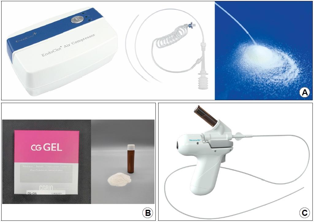
Hemostatic powders are commercially and clinically available in Korea. (A) Endoclot, (B) CGGEL, (C) Nexpowder.

The endoscopic views show the following. (A) A slightly elevated early gastric cancer with central ulceration at the antrum. (B) A post-resection ulcer with a maximum diameter of 45 mm after completion of endoscopic submucosal dissection. (C) The appearance after application of polysaccharide hemostatic powder to surface of the post-resection ulcer.
CONCLUSIONS
Although ESD is an excellent method for the treatment of gastric neoplasms, the appropriate strategy for the prevention and management of post-ESD adverse events is a problem still awaiting a solution. In particular, reducing post-ESD bleeding in high-risk patients is becoming increasingly important. Until now, no existing method can completely prevent post-ESD bleeding. Endoscopists need to always consider the risk of post-ESD bleeding and make maximum efforts to completely avoid this adverse event. Future studies should aim at establishing various methods and materials that are easier to use and have maximal preventive effects against post-ESD bleeding.
Notes
No potential conflict of interest relevant to this article was reported.