Korean J Helicobacter  Up Gastrointest Res Search

CLOSE


Korean J Helicobacter  Up Gastrointest Res > Volume 19(2); 2019 > Article
Kim: A Longstanding Mucosal Break in the Lower Esophagus?
Question: A 74-year-old man visited for the evaluation of indigestion for indigestion. Upper gastrointestinal endoscopy revealed a reddish longitudinal mucosal break at the lower esophagus; grade B reflux esophagitis (Los Angeles classification) was diagnosed (Fig. 1A). He was administered a proton pump inhibitor (PPI) once a day for 2 months. On follow-up endoscopies 2 and 3 years later, the mucosal break was still present (Fig. 1B, C), and then he took a PPI repeatedly. On follow-up endoscopy 4 years later, there was no change in the mucosal break (Fig. 1D).
What is the most likely diagnosis?
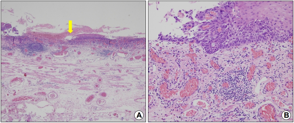
Answer: Endoscopic forceps biopsy was performed for the persistent mucosal break. Biopsy of this lesion disclosed a well-differentiated squamous cell carcinoma. EUS revealed tumor invasion of only the mucosa, and chest and abdominal CT scanning found no metastases. Therefore, he underwent endoscopic submucosal dissection that removed the lesion completely (Fig. 2). On histopathologic examination, the cancer cells were located at the falling off epithelial layer (Fig. 3A), and invasion of the cancer from the basal layer into the lamina propria mucosae was noted (Fig. 3B). This patient has been followed-up for 4 years since endoscopic submucosal dissection, with no evidence of recurrence.
Reflux esophagitis can progress to adenocarcinoma, as like many other chronic inflammatory conditions [1]. Inflammation in the mucosal break also can result in regenerative atypia that is sometimes confused with dysplasia, but that does not have malignancy as its end result [1]. Therefore, it is recommended to perform endoscopic biopsy when the inflammatory reaction has subsided after PPI administration. However, in the present case, the mucosal break was single, and its shape was not changed during follow-up. At this situation, endoscopic biopsy for the unchanged mucosal break is needed not to miss carcinoma mimicking inflammation [2]. We always remember that we should not make definite assumptions about histology from the gross appearance of a lesion.

CONFLICTS OF INTEREST

No potential conflict of interest relevant to this article was reported.

Fig. 1.
(A) Upper gastrointestinal endoscopy image showing a reddish longitudinal mucosal break in the lower esophagus, indicative of grade B reflux esophagitis (Los Angeles classification). (B~D) Follow-up endoscopy images at 2, 3, and 4 years later, showing a reddish longitudinal mucosal break in the lower esophagus.
kjhugr-2019-19-2-137f1.jpg
Fig. 2.
Endoscopic submucosal dissection. (A) A reddish mucosal break-like lesion in the lower esophagus. (B) Marking dots to demarcate the lesion. (C) A complete circumferential incision made using a hook knife. (D) Submucosal dissection made using an insulated-tip knife. (E) Complete removal of the lesion. (F) The resected specimen.
kjhugr-2019-19-2-137f2.jpg
Fig. 3.
(A) Histopathological examination of the cancer cells at the falling-off epithelial layer (arrow) (H&E, original magnification, ×40). (B) Magnification showing invasion of the cancer from the basal layer into the lamina propria mucosae (H&E, original magnification, ×400).
kjhugr-2019-19-2-137f3.jpg

REFERENCES

1. Kavanagh ME, O'Sullivan KE, O'Hanlon C, O'Sullivan JN, Lysaght J, Reynolds JV. The esophagitis to adenocarcinoma sequence; the role of inflammation. Cancer Lett 2014;345:182–189.
crossref pmid
2. Hosaka H, Kawamura O, Kusano M, Shimoyama Y, Fujimori T, Kuwano H. Early esophageal squamous cell carcinoma mimicking reflux esophagitis. Gastrointest Endosc 2010;71:1063–1064.
crossref pmid
TOOLS
Share :
Facebook Twitter Linked In Google+ Line it
METRICS Graph View
  • 0 Crossref
  •    
  • 3,621 View
  • 46 Download
Related articles in Korean J Helicobacter Up Gastrointest Res


Editorial Office
Lotte Gold Rose II Room 917, 31 Seolleung-ro 86-gil, Gangnam-gu, Seoul 06193, Korea
Tel: +82-2-717-5543    Fax: +82-2-565-9947    E-mail: helicojournal@smileml.com                

Copyright © 2024 by Korean College of Helicobacter and Upper Gastrointestinal Research.

Developed in M2PI

Close layer
prev next