오버튜브 삽입 후 발생한 식도 천공에서 내시경적 음압 치료로 치료한 1예
Endoscopic Vacuum-assisted Closure in a Patient with an Overtube-induced Esophageal Perforation
Article information
Trans Abstract
An esophageal perforation is one of the most fatal clinical events, with a mortality rate of up to 21%. This may arise postoperatively or post-endoscopically. In the past, surgical treatment, such as an esophagectomy, was performed these cases. However, the procedure was challenging and had a high risk of postoperative complications. Recently, advancements in endoscopic techniques have been made, and endoscopic procedures became a common treatment modality for patients with esophageal perforation, even in those with underlying diseases. Among the endoscopic procedures, endoscopic vacuum-assisted closure (E-VAC) has been known to be safe and effective. We present the case of a 64-year-old female with advanced liver cirrhosis and an overtube-induced esophageal perforation during esophageal variceal ligation. She was successfully treated with E-VAC.
서 론
식도 천공은 식도 절제술, 위 절제술 등 수술 이후 발생하는 문합부 유출 또는 내시경 등 시술 후 발생하는 의인성 천공, 또는 매우 심한 구역 및 구토 후 발생하는 부르하버 증후군의 형태로 나타날 수 있다[1,2]. 식도 천공은 약 10~21%의 사망률을 보이는 심각한 임상 증상으로, 발견되는 즉시 이에 대한 즉각적인 치료가 필요하며 치료가 지연될수록 환자의 사망률은 비약적으로 증가하게 된다[3-5]. 과거 식도 천공을 치료하기 위해서 식도 절제술 등의 수술적 치료를 시행하였으나, 수술적 치료 자체의 난이도 및 수술 후 합병증 발생의 위험률을 고려하였을 때 최근에는 비침습적 치료, 특히 내시경 치료를 통한 식도 천공의 치료가 다양한 방법으로 시도되고 있다. 그중 최근 여러 연구를 통해 endoscopic vacuum-assisted closure (E-VAC)가 식도 천공의 치료에 유용한 것으로 확인되었고, 기존에 사용되던 스텐트 삽입술과 비교하였을 때 치료 효과가 유의하게 좋은 것으로 나타났다[6,7]. E-VAC는 개흉, 개복 등의 침습적 조치를 요하지 않는 치료법으로, 특히 기저 질환 등으로 수술적 치료가 어려운 환자들에게서 유용하게 사용되고 있고, 경험이 축적되고 새로운 방법들이 개발되면서 더 많은 적응증에서 이용될 수 있을 것으로 기대된다. 본원에서 내시경 시술 도중 발생한 식도 천공 환자에서 E-VAC를 이용하여 성공적으로 식도 천공을 치료하였기에, 이에 대해 보고하고자 한다.
증 례
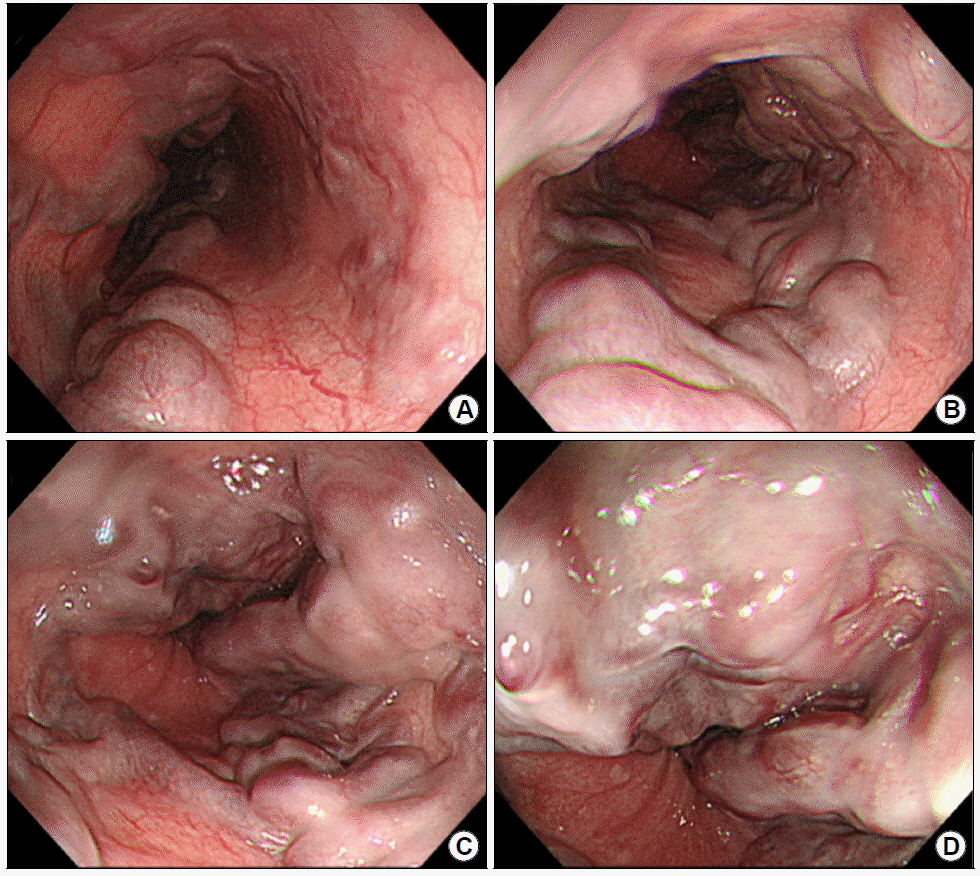
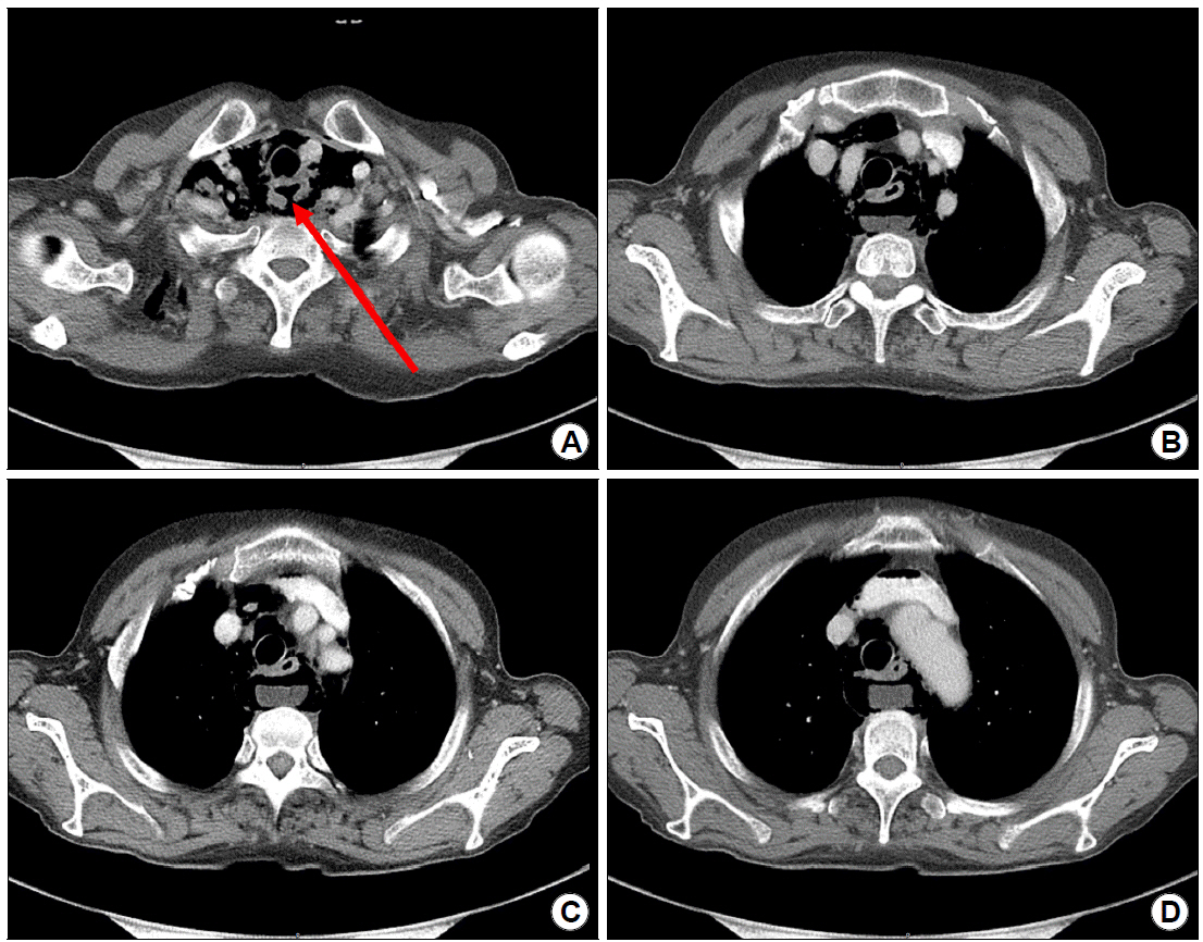
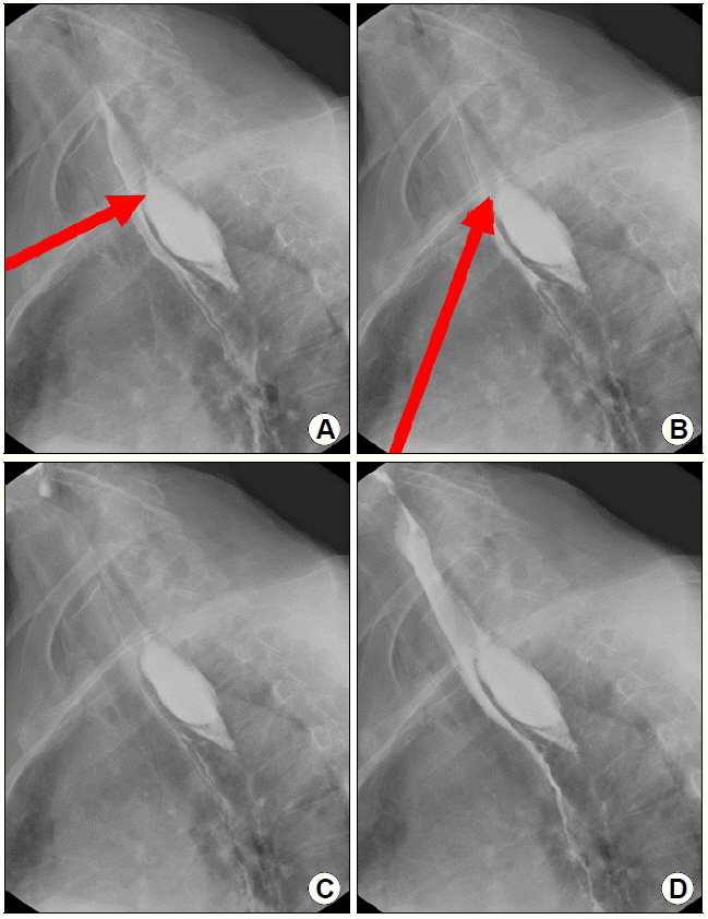
64세 여자 환자로 내시경 시술 도중 발생한 식도 천공으로 소화기내과 상부위장관 파트로 의뢰되었다. 환자는 2년 전 타원에서 진단된 자가면역 간염 및 이로 인한 간경화에 대한 추가 검사 및 치료 계획 수립을 위해 본원 입원하였다. 입원하여 시행한 위 내시경상 F3CbLs의 식도 정맥류 및 적색 소견 양성이 확인되어 입원 9일째 식도 정맥류 결찰술을 시행하기로 하였다(Fig. 1). 환자의 식도 정맥류의 범위가 광범위하여 여러 개의 밴드를 사용해야 할 것으로 생각되어 환자의 불편감 경감 및 시술의 편의성을 위해 먼저 오버튜브를 거치하기로 결정되었다. 상부위장관 내시경을 시행하기 전 오버튜브를 삽입하였으나 이물감으로 인해 환자가 지속적으로 기침을 하는 과정에서 오버튜브가 식도에서 밀려 나와 제거하였고, 제거한 후 상부위장관 내시경을 다시 삽입하고 관찰하였을 때 상부 식도 괄약근 직하방의 식도 점막에 깊은 열상이 확인되어 검사를 종료하였다. 증상 발생 직후 시행한 흉부 전산화단층촬영에서 상절치에서 16 cm 위치에 장경 약 2.8 cm 크기의 식도 천공이 관찰되었고(Fig. 2), 증상 발생 다음 날 시행한 식도조영술 검사에서도 흉곽입구 위치의 경부 식도에서 후벽의 전층 천공 및 종격동으로의 조영제 유출이 확인되었다(Fig. 3). 환자의 활력징후는 안정적이었으며 발열은 관찰되지 않았다. 혈액 검사상 백혈구 5,000/uL (다핵구 82.9%), C반응성 단백질 12.15 mg/dL의 염증이 동반되어 있었고, 혈소판 63,000/m3, 프로트롬빈 시간 76.0%의 응고장애가 확인되었다. 흉부외과에 수술적 처치에 대해 상의하였고 수술적 치료의 위험성이 높을 것으로 생각되어, 금식 및 경험적 항생제를 사용하면서 E-VAC를 시도하기로 하였다. 천공 발생 이틀 뒤 본과에 E-VAC가 의뢰되어 병변 확인을 위해 상부위장관 내시경을 시행하였고, 상절치에서 15~18 cm 위치에 점막 궤양 및 식도 천공 부위가 확인되어 E-VAC를 진행하였다. 본원에서는 미리 제조된 VAC kit (CuraVAC®; CGbio Inc., Sungnam, Korea)를 이용하였는데, 이 kit 내에는 의료용 폴리우레탄 스폰지, 봉합용 필름, 무균적으로 포장된 연결 튜브가 포함되어 있다. 먼저 상부위장관 내시경을 시행하여 천공이 발생한 위치를 확인한 뒤 내시경을 회수하고, 비강으로 16 Fr 비위관(2207-016; Sewoon Medical Co., Cheonan, Korea)을 삽입한 뒤 구강 밖으로 비위관을 회수하였다. 회수한 비위관 끝에 천공 크기에 맞추어 모양을 다듬은 폴리우레탄 스폰지를 나일론 또는 실크 등의 실로 단단히 꿰매어 고정한 뒤이를 겸자를 이용하여 상부 식도 괄약근을 지나 조심스럽게 삽입하였다. 내시경과 겸자를 조심스럽게 삽입하며 천공 부위로 접근하였으나 병변의 위치가 상부 식도 괄약근의 직하방으로, 천공 내부로 스폰지를 삽입하기에는(intracavitary) 접근이 쉽지 않아 천공의 바로 옆 식도 점막에 최대한 밀착하여 비위관과 스폰지를 위치시킨 뒤(intraluminal) 조심스럽게 내시경을 회수하였다(Fig. 4). 비위관이 빠지지 않도록 단단히 피부에 고정한 후 비위관의 한 쪽 끝에 100 mmHg의 음압을 적용한 진공 펌프를 적용하였다. 천공 발생 5일 뒤 첫 번째 추적 위 내시경을 시행하였고, 이전 위 내시경 소견과 비교하여 천공의 크기는 감소하고 천공 부위 주변의 궤양이 이전보다 호전된 양상이 확인되었다(Fig. 5A). 일반적으로는 천공의 크기가 변할 경우 천공의 크기에 맞추어 스폰지의 크기를 변경하여 E-VAC를 교체하는 경우가 많으나, 본 환자의 경우 intraluminal approach로 E-VAC를 거치한 상태로, 병변의 크기가 이전 내시경에 비해 확연히 감소하여 E-VAC가 원활하게 작용하고 있는 것으로 생각되었으며, intracavitary approach가 아니어서 스폰지의 크기를 변경하지 않아도 E-VAC의 기능이 원활할 것으로 생각되어 위 내시경 선단 및 겸자를 이용하여 스폰지의 위치를 조절하였다. 또한 천공 주변으로 다량의 액체 분비물이 고여있어 창상 부위의 건조를 유지하기 위해 음압을 120 mmHg로 높여 유지하였다. 이후 천공 발생 14일 뒤 두 번째 추적 위 내시경에서 천공은 치유되어 관찰되지 않았고 주변부 궤양 역시 회복기로 관찰되어 E-VAC를 제거하였다. E-VAC를 제거한 다음 날 추적 식도조영술을 시행하였고 마찬가지로 조영제 유출은 관찰되지 않아 천공이 폐쇄되었음을 확인하였다(Fig, 5B, C). 이후 환자는 조심스럽게 식이 진행을 단계적으로 시행하였고, 천공 발생 20일 후 급성 합병증 없이 퇴원하였다.

Initial endoscopic findings of the patient. (A, B) Several tortuous venous distensions may be seen in the esophagus. (C, D) Red color signs may be found in the lower esophagus, just above the gastroesophageal junction.

Chest CT images of the patient after perforation. (A) A 2.8 cm sized transmural esophageal wall defect may be seen in thoracic inlet level (arrow). (B-D) Loculated air-fluid collection in retropharyngeal space communicating with esophageal lumen due to esophageal defect, and extensive pneumomediastinum along esophagus may be found.

Esophagography images of the patient after perforation. (A, B) Transmural perforation in the posterior wall of the esophagus in the thoracic inlet level may be noted (arrow). (C, D) Loculated extraluminal leakage of the contrast media into the mediastinum was may be found.

Endoscopic images of endoscopic vacuum-assisted (E-VAC) closure procedure. (A) A 2 cm sized transmural perforation may be found found in UI 15~18 cm. (B) Polyurethane sponge is grasped with endoscopic forceps, and is inserted through the oral cavity. (C, D) Nasogastric tube with a polyurethane sponge is located near the perforation site. UI, upper incisor.

Follow-up endoscopic images and esophagography image. (A) A follow-up endoscopy performed 5 days after the E-VAC placement shows that the size of the perforation hole decreased, and ulceration of the peripheral mucosa also showed improvement. (B) A second follow-up endoscopy performed 7 days after the first follow-up endoscopy shows that the esophageal mucosa is in the healing stage. No residual perforation hole is noted. (C) Follow-up esophagography shows no extraluminal contrast leakage. E-VAC, endoscopic vacuum-assisted closure.
고 찰
오버튜브로 인한 점막 손상 및 이로 인한 경부 식도의 천공에 대해, 기저 질환으로 수술적 치료가 어려운 환자에서 E-VAC를 이용하여 합병증 없이 안전하게 치료할 수 있었다. E-VAC를 적용하여 치료하는 동안 환자는 발열, 흉통 및 패혈증을 의심할 증상 없이 치료를 진행하는 20일간 안정적인 경과를 보였고, 천공이 치료된 후에는 단계적 식이 진행까지 원활한 가운데 퇴원하였다.
식도 천공의 적절한 치료 방법을 결정하기 위해서 환자의 임상적인 상태뿐만 아니라 천공이 발생한 원인, 천공이 발생한 식도 위치 및 천공 부위의 크기 등이 함께 고려되어야 한다[4,8]. 먼저 흉부 X-ray, 흉부 전산화단층촬영 및 바륨 등을 이용한 식도조영술 등의 검사를 통해 정확한 식도 천공의 위치 및 범위에 대해 평가한 후, 감염이 조절되지 않거나 이로 인한 쇼크, 호흡 부전 등 불안정한 활력징후가 보일 경우, 또는 종격동 기종이 넓은 범위에서 관찰되거나 기흉, 기복증 등이 동반되었을 경우에는 조속히 수술적 처치를 고려할 것이 권고된다[4,8,9]. 본 환자에서는 여러 영상학적 검사에서 명확한 식도 천공 소견이 확인되었고 혈액 검사에서 급성 염증 소견이 있었으나, 환자의 기저 간질환으로 인한 응고 장애가 동반되어 있어 수술적 치료 후 발생할 출혈 등의 합병증 발생을 고려하였을 때 수술적 치료를 우선적으로 시행하기에는 적합하지 않을 것으로 생각되었고, 비수술적 치료를 진행하는 동안에도 광범위 항생제 및 E-VAC를 유지하면서 염증이 잘 조절되는 경과를 보여 응급 수술을 요하는 상황은 초래되지 않았다.
최근 들어 식도 천공이 발생한 환자에서 내시경적 클립 봉합술, 내시경적 스텐트 삽입술 및 본 환자와 같이 E-VAC 등의 내시경적 시술을 통한 비침습적 치료를 우선적으로 시도하고 있다. 내시경적 클립 봉합술은 1993년 Binmoeller 등[10]이 위 평활근종을 절제한 후 내시경 클립을 이용하여 봉합한 예를 발표하면서 처음 소개되었다. 내시경적 클립 봉합술은 봉합을 발견한 즉시 시도할 수 있다는 장점이 있으며, 특히 병변의 크기가 1~2 cm가량의 비교적 그 천공의 크기가 작은 경우에 효과적이다[11,12]. 그러나 본 환자와 같이 천공의 크기가 2 cm 이상일 경우는 기술적으로 점막 및 점막하층의 봉합이 어려워 결과적으로 식도 천공의 치료에 제한적인 적응증을 보였다.
클립을 이용하여 봉합하기 어려운 비교적 크기가 큰 식도 천공에서는 스텐트를 이용하여 봉합을 시도하기도 하며, 특히 2017년 Persson 등[13]의 연구 결과에 따르면 식도 수술 후 문합부 누출이 있는 환자에서 스텐트 삽입군의 치료 효과(88%)가 수술군의 치료 효과(83%)와 비교하여 열등하지 않으며 합병증 발생률은 스텐트 삽입군(7.5%)이 수술군(17%)보다 낮아 수술을 대체할 수 있는 효과적이고 안전한 치료 방법임을 입증한 바 있다. 그러나 스텐트를 장기간 거치할 경우 스텐트의 이동 및 제거, 통증이 발생할 수 있으며, 또한 스텐트가 식도 내강에 지속적으로 자극을 줌으로써 식도 궤양이 발생하거나 식도 협착이 발생하는 등 합병증 발생의 위험성이 증가하고, 합병증이 발생할 때마다 스텐트를 주기적으로 교체하거나 제거해야 한다는 불편감이 있다[14,15]. 본 환자의 경우 병변이 상부 식도 괄약근 직하방에 위치하고 있어 스텐트가 위치할 수 있는 공간이 부족하여 스텐트를 거치하기 어려웠으며, 스텐트로 인한 자극으로 심한 통증이 유발될 수 있어 적절하지 않은 치료 방법으로 생각되었다.
Vacuum treatment는 1993년 Fleischmann 등[16]이 개방성 골절을 입은 환자의 감염성 창상에 사용한 후 처음 보고되었고, 2008년 Wedemeyer 등[17]이 식도 수술 후 문합부 누출이 발생한 환자에서 내시경적으로 처음 적용하여 보고한 이후 다양한 부위에서 사용되고 있다. 국내에서도 여러 논문을 통하여 식도암 수술 후 발생한 문합부 누출에 E-VAC를 적용함으로써 약 70~95%의 높은 치료 성공률을 보였음을 확인한 바 있다[18,19]. 또한 여러 연구에서 식도 천공이 발생한 환자에 대해 식도 스텐트를 삽입한 환자와 E-VAC를 시행한 환자를 비교하였을 때 치료 기간에는 유의한 차이가 없으면서 천공 폐쇄율이 더 유의하게 높았고 사망률은 유의하게 낮아 그 유효성과 안전성을 확인하였다[6,7,20]. 다만 E-VAC를 시행하기 전 몇 가지 고려해야 할 점이 있다. 먼저 스텐트 삽입군과 비교하여 E-VAC군에서 스텐트군에 비해 내시경을 반복하는 횟수가 유의하게 많았는데, 이와 같이 반복적인 내시경 검사로 인해 환자들의 불편감이 증가할 수 있다[20]. E-VAC를 적용한 환자에서 내시경을 반복하는 정확한 기간 및 횟수는 아직까지 명확히 정해진 바는 없다. 본원에서는 시술과 관련된 환자들의 불편감을 최소화하기 위해 약 1주일 간격으로 추적 내시경을 시행한 후 E-VAC의 위치 또는 음압을 조절하거나 제거 여부를 결정하고 있으며, 본 환자에서도 마찬가지로 5~7일 간격으로 추적 내시경을 시행하여 병변의 호전 여부를 확인하였다. 또한 구강 내로 삽입 가능한 스폰지의 크기에 제한이 있기 때문에 실질적으로 천공의 크기가 4 cm 이상의 큰 병변의 경우에는 E-VAC를 시행하기에 어려움이 있다. 또한 병변의 위치 및 특성에 따라 분비물이 상대적으로 많은 기관-위 샛길의 경우 진공 펌프만으로 충분히 분비물을 제거하기 힘들어 E-VAC로 충분한 효과를 보이기 어려울 수 있고, 병변의 위치가 연동운동이 활발한 부분에 위치하고 있을 때는 E-VAC가 이동할 가능성이 높기 때문에 효과가 제한적일 수 있다[18]. 따라서 환자의 임상적인 상황 및 병변의 특징을 종합적으로 고려하여 치료 방법을 결정하는 것이 필요할 것으로 고려된다.
정리하자면, 식도 천공이 발생한 환자에서 수술적 치료보다 내시경 치료를 통한 비침습적 치료가 우선적으로 시행되고 있다. 그중 E-VAC는 기저 질환 등으로 수술적 치료가 어려운 환자에서 다른 비침습적 내시경 시술들과 비교하였을 때 치료 효과가 높고 상대적으로 합병증 발생이 적어 안전하게 사용될 수 있어, 비침습적 치료를 시행할 때 선택할 수 있는 하나의 치료적 대안이 될 수 있을 것으로 기대된다.
Notes
No potential conflict of interest relevant to this article was reported.
