Korean J Helicobacter  Up Gastrointest Res Search

CLOSE


Korean J Helicobacter  Up Gastrointest Res > Volume 22(4); 2022 > Article
내시경 점막하 박리술로 진단된 상피하종양
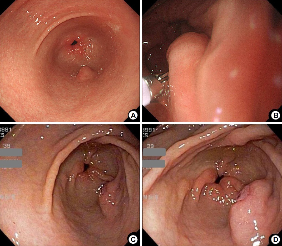
요약: 당뇨, 고지혈증의 기저 병력을 가진 41세 남자가 검진 목적으로 상부위장관 내시경 검사를 시행받았다. 내시경 검사에서 약 1 cm 가량의 상피하종양이 전정부 대만 부위에서 발견되었으며, 생겸 겸자로 눌러봤을 때 rolling sign 양성, cushion sign 양성이었다(Fig. 1A, B). 상피하종양의 표면에서 겸자로 조직검사를 시행하였다. 조직 검사 결과는 만성위염으로 보고되었으며 장상피화생과 헬리코박터 감염은 동반되지 않았다.
후향적으로 과거 상부위장관 내시경 검사를 검토해 보았고, 내원 14개월 전 다른 병원에서 시행한 상부위장관 내시경에서 중심부에 미란을 동반한 상피하종양 병변이 같은 부위에서 관찰되었다(Fig. 1C, D). 이번에 시행한 내시경 소견을 비교하였을 때, 상피하종양의 크기 감소 및 중심부 미란의 소실이 확인되었다.
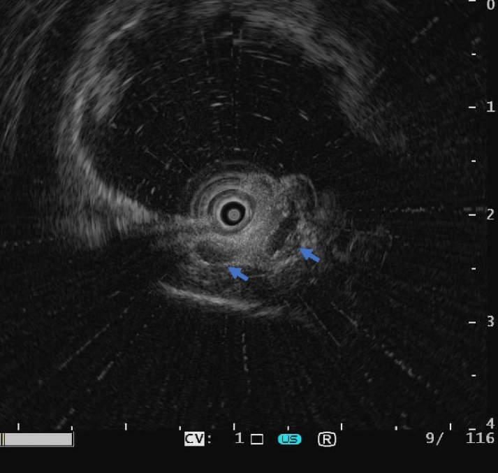
상피하종양에 대한 추가 평가를 위해 내시경 초음파 검사(endoscopic ultrasound)를 시행하였다(Fig. 2). 내시경 초음파 검사에서 점막하층에 불균질한 저에코를 가진 13 mm 정도의 병변이 관찰되었으며, 병변 내부에 다발성 낭성 구조가 포함되어 있었다. 주변 구조와 경계는 명확하였다.
본원에서 시행한 내시경에서는 상피하종양의 표면점막에서 미란은 관찰되지 않았으나, 14개월 전 타원에서 시행한 내시경 소견에서 상피하종양 표면 점막에 미란이 존재하였기 때문에 정확한 진단을 위해 내시경 점막하 박리술을 시행하였다(Fig. 3). 상기 환자 상피하종양의 원인으로 가능성이 높은 질환은?
해설: 심재성 낭종성 위염(gastritis cystica profunda)은 양성 위샘의 낭종성 확장과 점막하 침윤을 특징으로 하는 비교적 드문 질환으로[1], 1972년 위 수술 후 문합부의 용종 및 종물형 병변을 평가하는 도중 최초로 증례보고된 질환이다[2]. 초기에는 위 수술 병력이 있는 경우에 발생하는 것으로 보고되었지만[2], 현재는 위 수술력 없이 심재성 낭종성 위염이 발견된 다양한 증례들이 다수 보고되었기 때문에 수술 과거력과 심재성 낭종성 위염의 발생은 직접적인 연관은 없는 것으로 판단된다. 질병의 임상적 특성에 대해 알려진 내용이 많지 않으나, 한 국내 연구에서는 60대에서 호발하며, 남성에서 많이 생기며, 전정부와 체부에서 많이 발생한다고 하였다[3]. 다른 해외 연구에서도 대부분 50대 이상에서 발생하며, 여성보다는 남성에서 많이 생긴다고 보고하였고, 호발 부위는 체부, 궁륭부, 전정부 순으로 보고하였다[4].
심재성 낭종성 위염의 임상적인 의미를 고찰해 보자면, 과거에는 수술 문합부 주변의 가성종양으로 발견되어 악성 종양의 국소재발과 감별이 필요한 병변이었다면, 현재에는 내시경적으로 우연히 발견되는 용종 또는 상피하종양으로 내시경 육안 소견과 겸자를 이용한 조직 검사로는 진단이 쉽지 않기 때문에 내시경 점막하 박리술이나 수술 등을 하고 나서야 진단이 되는 병변이라는 점이다.
심재성 낭종성 위염의 임상 증상은 폐색, 출혈을 동반하는 경우부터 내시경적 검사에서 우연히 발견되는 경우까지 다양하게 보고되고 있으며[2,4-7], 용종이나 상피하종양으로 주로 발견된다는 점을 고려할 때 비특이적인 소화기 증상 평가를 위한 상부위장관 내시경 검사에서 우연히 발견되는 경우가 점차 증가하고 있다.
심재성 낭종성 위염의 내시경적 육안 소견은 매우 다양한 것으로 알려져 있다. 용종 형태나 상피하종양 형태가 비교적 흔하며, 드문 형태로는 거대주름 형태로도 나타나는 경우도 보고되어 있다. 내시경 초음파 검사 소견은 점막하층에 다발성의 무에코성의 낭성 병변을 가진 경우가 가장 흔히 발견되는 소견이다[8].
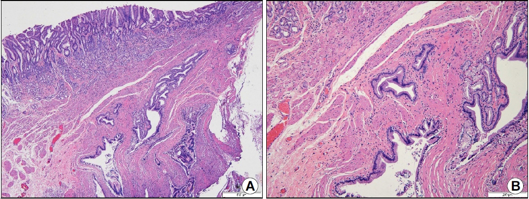
본 증례에서도 내시경 초음파를 시행했을 당시에는 이소성 췌장을 우선 의심하였었다. 그렇지만 상피하종양 표면점막의 변화가 있었던 상태라 정확한 진단을 위하여 내시경 점막하 박리술을 시행하였으며, 최종 조직병리 보고상 낭성으로 확장된 위샘이 점막하층과 고유근층을 침범하고 있어 심재성 낭종성 위염으로 진단되었다(Fig. 4). 이형성(dysplasia)은 관찰되지 않았으며, 절제 변연의 침범 소견도 관찰되지 않았다. 추적 관찰 방법에 있어서 아직 정립된 내용은 없으나, 본 증례에서는 1년 후 추적 관찰 내시경을 시행하기로 하였다.
심재성 낭종성 위염의 경우 일반적으로 양성 경과를 가진다고 알려져 있으나, 심재성 낭종성 위염이 전암성 병변 또는 암주위 병변일 가능성을 시사하는 보고가 있다. 특히 심재성 낭종성 위염이 위치한 표층 점막에서 악성 종양이 발견된 증례보고도 있으며[9], 최근에는 심재성 낭종성 위염 발생에 분자유전학적인 이상이 동반되었을 가능성에 관한 보고들이 나오고 있어서[10], 심재성 낭종성 위염 환자들을 추적 관찰할 때 다소 주의가 필요할 것으로 생각이 된다.
치료 방법이 아직 명확히 정립되어 있는 것은 아니지만, 출혈이나 위장관 폐쇄가 있으면 수술적 치료를 고려해 봐야 할 것이며, 무증상이더라도 악성 질환의 배제 등이 필요하다면 내시경 점막하 박리술을 통한 진단 및 치료를 적극 고려해 봐야 할 것이다. 현재까지 심재성 낭종성 위염 관련 대부분 증례에서 수술 또는 내시경 점막하 박리술을 시행하여 최종 진단되었다는 점을 고려한다면, 당분간은 수술 또는 내시경적 절제가 주된 치료 방침이 될 것으로 예상이 되나, 표면점막의 변화가 없으며, 내시경 초음파 검사상 심재성 낭종성 위염이 명확하다면 주의 깊은 추적 관찰도 하나의 대안이 될 수 있을 것으로 보인다.

CONFLICTS OF INTEREST

No potential conflict of interest relevant to this article was reported.

Fig. 1.
Endoscopic findings. (A) Image showing an antral subepithelial tumor (SET) measuring 1 cm in size. (B) The SET appears soft in consistency with a positive cushion sign. (C) Image (distant view) obtained at a previous hospital, 14 months before the patient visited our center. (D) Image (close-up view) showing an erosion on the surface of the SET.
kjhugr-2022-0041f1.jpg
Fig. 2.
Endoscopic ultrasound finding. A subepithelial tumor (13 mm) originating in the submucosal layer is observed. The inhomogeneous hypoechoic mass contains multiple cystic components (arrows).
kjhugr-2022-0041f2.jpg
Fig. 3.
Images showing endoscopic submucosal dissection with en-bloc resection of the subepithelial tumor.
kjhugr-2022-0041f3.jpg
Fig. 4.
Histopathological findings. (A) Image showing the mucosa, submucosa, and a part of the muscularis propria layer (from the surface inward). Cystic dilatation of gastric glands that penetrate the submucosa and muscularis propria is visualized beneath the normal-appearing mucosa (Hematoxylin and Eosin [H&E] stain, ×40). (B) Image (magnified view) showing normal gastric mucosa without dysplastic change in the muscularis propria layer (H&E stain, ×100).
kjhugr-2022-0041f4.jpg

REFERENCES

1. Koga S, Watanabe H, Enjoji M. Stomal polypoid hypertrophic gastritis: a polypoid gastric lesion at gastroenterostomy site. Cancer 1979;43:647–657.
crossref pmid
2. Littler ER, Gleibermann E. Gastritis cystica polyposa. Gastric mucosal prolapse at gastroenterostomy site, with cystic and infiltrative epithelial hyperplasia. Cancer 1972;29:205–209.
crossref pmid
3. Lee HJ, Lee TH, Lee JU, et al. Clinical features of gastritis cystica profunda in patients without history of gastric surgery (gastric cancer patients vs. non-cancerous patients). Korean J Med 2006;71:511–517.

4. Du Y, Zhang W, Ma Y, Qiu Z. Gastritis cystica profunda: a case report and literature review. Ann Palliat Med 2020;9:3668–3677.
crossref pmid
5. Yoon JE, Kim MS, Lee KC, Park HJ, Park CI. A case of gastritis cystica profunda with a long stalk presenting with upper gastrointestinal bleeding. Korean J Gastrointest Endosc 2007;35:186–189.

6. Kurland J, DuBois S, Behling C, Savides T. Severe upper-GI bleed caused by gastritis cystica profunda. Gastrointest Endosc 2006;63:716–717.
crossref pmid
7. Lee JH, Lee GS, Lee ES, et al. A case of gastritis cystica profunda presenting with a submucosal cystic tumor. Korean J Gastrointest Endosc 2008;37:280–283.

8. Machicado J, Shroff J, Quesada A, et al. Gastritis cystica profunda: endoscopic ultrasound findings and review of the literature. Endosc Ultrasound 2014;3:131–134.
crossref pmid pmc
9. Kim W, Park SC, Lee JY, et al. A case of gastric adenocarcinoma diagnosed after being followed up as submucosal tumor for 10 years. Korean J Gastroenterol 2001;37:291–295.

10. Itami H, Morita K, Nakai T, et al. Gastritis cystica profunda is associated with aberrant p53 and Epstein-Barr virus in gastric cancer: a clinicopathological, immunohistochemical and in situ hybridization study. Pathol Int 2021;71:42–50.
crossref pmid pdf


Editorial Office
Lotte Gold Rose II Room 917, 31 Seolleung-ro 86-gil, Gangnam-gu, Seoul 06193, Korea
Tel: +82-2-717-5543    Fax: +82-2-565-9947    E-mail: helicojournal@smileml.com                

Copyright © 2024 by Korean College of Helicobacter and Upper Gastrointestinal Research.

Developed in M2PI

Close layer
prev next