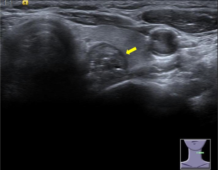
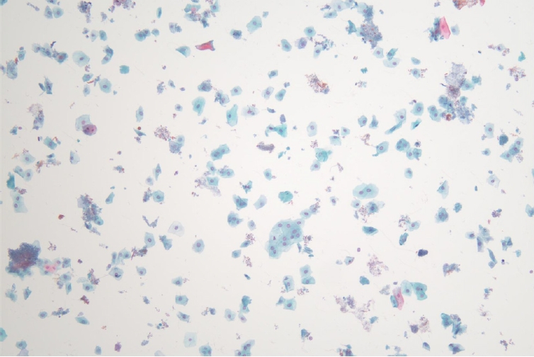
ņÜöņĢĮ: 62ņäĖ ņŚ¼ņ×ÉĻ░Ć Ļ▒┤Ļ░ĢĻ▓Ćņ¦äņŚÉņä£ ņŗżņŗ£ĒĢ£ Ļ░æņāüņāś ņ┤łņØīĒīī Ļ▓Ćņé¼ņŚÉņä£ ņÜ░ņŚ░Ē׳ ļ░£Ļ▓¼ļÉ£ Ļ▓░ņĀłļĪ£ ļé┤ņøÉĒĢśņśĆļŗż. ĻĖ░ņĀĆ ņ¦łĒÖśņØ┤ļéś ņé¼ĒÜīļĀź ļ░Å Ļ░ĆņĪ▒ļĀźņŚÉņä£ ĒŖ╣ņØ┤ ņåīĻ▓¼ņØĆ ņŚåņŚłņ£╝ļ®░, ņŗĀņ▓┤ Ļ▓Ćņé¼ņŚÉņä£ļÅä ņØ┤ņāü ņåīĻ▓¼ņØĆ Ļ┤Ćņ░░ļÉśņ¦Ć ņĢŖņĢśļŗż. Ļ▓Ćņ¦äņ£╝ļĪ£ ņŗ£Ē¢ēĒĢ£ Ļ░ĆņŖ┤ XņäĀ Ļ▓Ćņé¼ņÖĆ ņāüļČĆņ£äņןĻ┤Ć ļé┤ņŗ£Ļ▓Į Ļ▓Ćņé¼ņŚÉņä£ ņØ┤ņāü ņåīĻ▓¼ņØĆ ņŚåņŚłņ£╝ļ®░, Ļ░æņāüņāś ĻĖ░ļŖź Ļ▓Ćņé¼ļŖö T3 132.7 ng/dL, free T4 1.18 ng/dL, thyroid stimulating hormone 1.60 uIU/mLļĪ£ ņĀĢņāüņØ┤ņŚłļŗż. ļ│ĖņøÉņŚÉņä£ ņŗ£Ē¢ēĒĢ£ Ļ░æņāüņāś ņ┤łņØīĒīī Ļ▓Ćņé¼ņŚÉņä£ Ļ░æņāüņāśņØś ņóīņāüņŚĮņŚÉ ņĢĮ 1.3 cm ├Ś 1.2 cm Ēü¼ĻĖ░ņØś Ļ▓░ņĀłņØ┤ Ļ┤Ćņ░░ļÉśņŚłņ£╝ļ®░(Fig. 1), ņØ┤ņŚÉ ļīĆĒĢ┤ ņäĖņ╣©ĒØĪņØĖ Ļ▓Ćņé¼ļź╝ ņŗ£Ē¢ēĒĢśņśĆļŗż. ņäĖĒż Ļ▓Ćņé¼ņŚÉņä£ ļŗżņłśņØś ĒÄĖĒÅēņäĖĒż(squamous cell) ņÖĖņŚÉ ĒŖ╣ņØ┤ ņåīĻ▓¼ņØĆ ņŚåņŚłļŗż(Fig. 2).
ļŗżņØīņŚÉ ņŗ£Ē¢ēĒĢĀ ņĀüņĀłĒĢ£ Ļ▓Ćņé¼ņÖĆ Ļ░ĆļŖźņä▒ņØ┤ ļåÆņØĆ ņ¦äļŗ©ņØĆ?
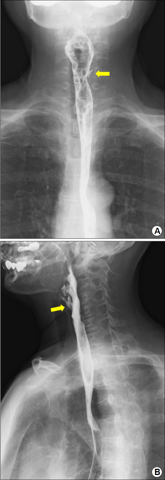
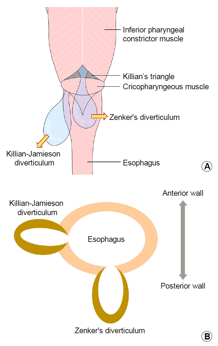
ĒĢ┤ņäż: ņäĖĒż Ļ▓Ćņé¼ņŚÉņä£ ļŗżņłśņØś ĒÄĖĒÅēņäĖĒżĻ░Ć Ļ┤Ćņ░░ļÉśņŚłņØīņØä ĒåĀļīĆļĪ£ ņŗØļÅä ļ│æļ│Ć ņØśņŗ¼ ĒĢśņŚÉ ņŗØļÅäņĪ░ņśüņłĀĻ│╝ ņāüļČĆņ£äņןĻ┤Ć ļé┤ņŗ£Ļ▓Į Ļ▓Ćņé¼ļź╝ Ļ│äĒÜŹĒĢśņśĆļŗż. ņŗØļÅäņĪ░ņśüņłĀņŚÉņä£ ļ░öļź©ņØ┤ ņāüļČĆņŗØļÅäņØś ņĀäļ░®, ņóīņĖĪņ£╝ļĪ£ ļÅīņČ£ļÉśļŖö ņ×æņØĆ Ļ▓īņŗżņØ┤ Ļ┤Ćņ░░ļÉśņŚłļŗż(Fig. 3). ņāüļČĆņ£äņןĻ┤Ć ļé┤ņŗ£Ļ▓Į Ļ▓Ćņé¼ ņŗ£ņŚÉļŖö ņāüļČĆņŗØļÅäļź╝ ņל Ļ┤Ćņ░░ĒĢśĻĖ░ ņ£äĒĢ┤ ņ║ĪņØä ļČĆņ░®ĒĢ£ ļÆż ņŗ£Ē¢ēĒĢśņśĆņ£╝ļ®░, ņāüļČĆņŗØļÅäņĪ░ņ×äĻĘ╝ ņ¦üĒĢśļ░®ņØś ņāüļČĆņŗØļÅäņØś ņóīņĖĪļ▓ĮņŚÉ ņØīņŗØļ¼╝ņØ┤ Ļ│ĀņŚ¼ ņ׳ļŖö Ļ▓īņŗżņØ┤ Ļ┤Ćņ░░ļÉśņŚłļŗż(Fig. 4). ņāüĻĖ░ ņåīĻ▓¼ņØä ĒåĀļīĆļĪ£ Killian-Jamieson Ļ▓īņŗżļĪ£ ņ¦äļŗ©ĒĢśņśĆļŗż. ĒÖśņ×ÉļŖö Ļ▓īņŗż ļé┤ ņØīņŗØļ¼╝ņØś ņĀĆļźśļĪ£ ņØĖĒĢ£ Ļ▓āņ£╝ļĪ£ ņāØĻ░üļÉśļŖö ĻĄ¼ņĘ© ņ”ØņāüņØ┤ ņ׳ņŚłņ¦Ćļ¦ī, ņé╝Ēé┤Ļ│żļ×Ć, ĻĄ¼ĒåĀ, ĻĖ░ņ╣©Ļ│╝ Ļ░ÖņØĆ ņ”ØņāüņØ┤ ņŚåņ¢┤ Ēśäņ×¼ Ļ▓ĮĻ│╝ Ļ┤Ćņ░░ ņżæņØ┤ļŗż.
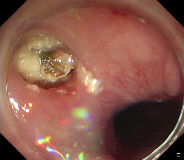
Killian-Jamieson Ļ▓īņŗżņØĆ ņ£żņāüņØĖļæÉĻĘ╝Ļ│╝ ņäĖļĪ£ņŗØļÅäĻĘ╝ ņé¼ņØ┤ņØś ņĘ©ņĢĮļČĆņ£äņŚÉņä£ Ļ▓ĮļČĆņŗØļÅä ņĀäņÖĖņĖĪņ£╝ļĪ£ ļÅīņČ£ļÉśņ¢┤ ņāØĻĖ░ļŖö ļ¦żņÜ░ ļō£ļ¼Ė Ļ▓īņŗżļĪ£, ņóģņóģ Zenker Ļ▓īņŗżĻ│╝ Ēś╝ļÅÖļÉśĻĖ░ ņēĮļŗż(Fig. 5A) [1]. ņŗØļÅäņĪ░ņśüņłĀĻ│╝ ļé┤ņŗ£Ļ▓Į Ļ▓Ćņé¼ ņŗ£ Zenker Ļ▓īņŗżņØĆ ņŗØļÅä Ēøäļ▓ĮņĖĪņŚÉņä£, Killian-Jamieson Ļ▓īņŗżņØĆ ņŗØļÅä ņĀäņÖĖņĖĪņŚÉņä£ Ļ┤Ćņ░░ļÉśļŖö Ļ▓āņØ┤ Ļ░Éļ│äņĀÉņØ┤ļŗż(Fig. 5B). ņāüļČĆņŗØļÅä Ļ▓īņŗżņØĆ Ļ░æņāüņāś Ēøäļ░®ņŚÉ ņØĖņĀæĒĢ┤ ņ׳ņ¢┤ Ļ░æņāüņāś ņ┤łņØīĒīī Ļ▓Ćņé¼ņŚÉņä£ ņóģņóģ Ļ░æņāüņāś Ļ▓░ņĀłļĪ£ ņśżņØĖļÉśĻĖ░Ļ░Ć ņē¼ņÜ░ļ®░, ļ│Ė ņ”ØļĪĆņÖĆ Ļ░ÖņØ┤ ļé┤ļČĆņŚÉ Ļ│ĀņŚÉņĮö ļČĆļČäņØä Ļ░Ćņ¦ĆļŖö ņøÉĒśĢņØ┤ļéś ĒāĆņøÉĒśĢņØś ņĀĆņŚÉņĮö ļ│æļ│Ćņ£╝ļĪ£ Ļ┤Ćņ░░ļÉ£ļŗż[2]. ļé┤ņŗ£Ļ▓Į Ļ▓Ćņé¼ ņŗ£ ņäĖņŗ¼ĒĢśĻ▓ī Ļ┤Ćņ░░ĒĢśņ¦Ć ņĢŖņ£╝ļ®┤ ņāüļČĆņŗØļÅäņĪ░ņ×äĻĘ╝ ņŻ╝ņ£äņØś Ļ▓īņŗżņØä ļåōņ╣Ā ņłś ņ׳ļŖöļŹ░, ļ│Ė ņ”ØļĪĆņŚÉņä£ļÅä ĒāĆ ļ│æņøÉņŚÉņä£ ņŗżņŗ£ĒĢ£ Ļ▓Ćņ¦ä ļé┤ņŗ£Ļ▓Į Ļ▓Ćņé¼ņŚÉņä£ļŖö Ļ▓īņŗżņØä ĒÖĢņØĖĒĢśņ¦Ć ļ¬╗ĒĢśņśĆļŗż. ņØ┤ļ¤¼ĒĢ£ Ļ▓ĮņÜ░ ņ╣śļŻīļé┤ņŗ£Ļ▓ĮņŚÉ ņé¼ņÜ®ļÉśļŖö ņ║ĪņØä ļČĆņ░®ĒĢśņŚ¼ Ļ▓Ćņé¼ļź╝ ņŗ£Ē¢ēĒĢśļ®┤ ļ╣äĻĄÉņĀü ņĢłņĀĢņĀüņ£╝ļĪ£ ņāüļČĆņŗØļÅäņĪ░ņ×äĻĘ╝Ļ│╝ ņāüļČĆņŗØļÅäļź╝ ņל Ļ┤Ćņ░░ĒĢĀ ņłś ņ׳ņ¢┤ ņ×æņØĆ Ļ▓īņŗżļ┐Éļ¦ī ņĢäļŗłļØ╝ ņóģņ¢æļÅä ļåōņ╣śņ¦Ć ņĢŖĻ│Ā Ļ▓Ćņé¼Ļ░Ć Ļ░ĆļŖźĒĢśļŗż.
Killian-Jamieson Ļ▓īņŗżņØĆ ļīĆļČĆļČä ļ¼┤ņ”Øņāüņ£╝ļĪ£ ņÜ░ņŚ░Ē׳ ļ░£Ļ▓¼ļÉśļ®░, ļ│┤ĒåĄ Ļ▓īņŗżņØś Ēü¼ĻĖ░Ļ░Ć 2.5 cm ņØ┤ņāüņØ┤ļ®┤ ņé╝Ēé┤Ļ│żļ×Ć, ņŚŁļźś, ņØĖļæÉĻĄ¼, ĻĖ░ņ╣©, ņē░ ļ¬®ņåīļ”¼, ĻĄ¼ņĘ© ņ”ØņāüņØä ņ£Āļ░£ĒĢĀ ņłś ņ׳ļŗż[3]. ņ”ØņāüņØ┤ ņ׳ļŖö Ļ▓ĮņÜ░ ņÖĖĻ│╝ņĀü Ļ▓īņŗżņĀłņĀ£ņłĀņØ┤ Ēæ£ņżĆ ņ╣śļŻīļĪ£ ņŗ£Ē¢ēļÉśņ¢┤ ņÖöņ£╝ļ®░, ņĄ£ĻĘ╝ņŚÉļŖö ļé┤ņŗ£Ļ▓ĮņĀü ņżæĻ▓®ņĀłĻ░£ņłĀ(endoscopic septostomy)ņØś ņ£ĀņÜ®ņä▒ņŚÉ ļīĆĒĢ£ ļ│┤Ļ│ĀĻ░Ć ņ”ØĻ░ĆĒĢśĻ│Ā ņ׳ļŗż[3].